The century of THE SURGEONS
Surgery is a profession defined by its authority to cure by means of bodily invasion. The brutality and risks of opening a living person’s body have long been apparent, the benefits only slowly and haltingly worked out. Nonetheless, over the past two centuries, surgery has become radically more effective, and its violence substantially reduced — changes that have proved central to the development of mankind’s abilities to heal the sick.
Consider, for instance, amputation of the leg.
The procedure had long been recognized as lifesaving, in particular for compound fractures and other wounds prone to sepsis, and at the same time horrific. Before the discovery of anesthesia, orderlies pinned the patient down while an assistant exerted pressure on the femoral artery or applied a tourniquet on the upper thigh.
Surgeons using the circular method proceeded through the limb in layers, taking a long curved knife in a circle through the skin first, then, a few inches higher up, through the muscle, and finally, with the assistant retracting the muscle to expose the bone a few inches higher still, taking an amputation saw smoothly through the bone so as not to leave splintered protrusions. Surgeons using the flap method, popularized by the British surgeon Robert Liston, stabbed through the skin and muscle close to the bone and cut swiftly through at an oblique angle on one side so as to leave a flap covering the stump.
The limits of patients’ tolerance for pain forced surgeons to choose slashing speed over precision. With either the flap method or the circular method, amputation could be accomplished in less than a minute, though the subsequent ligation of the severed blood vessels and suturing of the muscle and skin over the stump sometimes required 20 or 30 minutes when performed by less experienced surgeons.
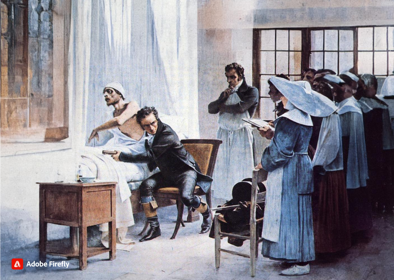
No matter how swiftly the amputation was performed, however, the suffering that patients experienced was terrible. Few were able to put it into words. Among those who did was Professor George Wilson. In 1843, he underwent a Syme amputation — ankle disarticulation — performed by the great surgeon James Syme himself. Four years later, when opponents of anesthetic agents attempted to dismiss them as “needless luxuries,” Wilson felt obliged to pen a description of his experience:
“The horror of great darkness, and the sense of desertion by God and man, bordering close on despair, which swept through my mind and overwhelmed my heart, I can never forget, however gladly I would do so. During the operation, in spite of the pain it occasioned, my senses were preternaturally acute, as I have been told they generally are in patients in such circumstances. I still recall with unwelcome vividness the spreading out of the instruments: the twisting of the tourniquet: the first incision: the fingering of the sawed bone: the sponge pressed on the flap: the tying of the blood-vessels: the stitching of the skin: the bloody dismembered limb lying on the floor.”
It would take a little while for surgeons to discover that the use of anesthesia allowed them time to be meticulous. Despite the advantages of anesthesia, Liston, like many other surgeons, proceeded in his usual lightning-quick and bloody way. Spectators in the operating-theater gallery would still get out their pocket watches to time him. The butler’s operation, for instance, took an astonishing 25 seconds from incision to wound closure. (Liston operated so fast that he once accidentally amputated an assistant’s fingers along with a patient’s leg, according to Hollingham. The patient and the assistant both died of sepsis, and a spectator reportedly died of shock, resulting in the only known procedure with a 300% mortality.)
The Surgical Personality

Surgical stereotypes are remnants of the days of pre-anaesthesia surgery and include impulsivity, narcissism, authoritativeness, decisiveness, and thinking hierarchically. Medical students hold these stereotypes of surgeons early in their medical training. As Pearl Katz says in the The Scalpel’s Edge: ‘Each generation perpetuates the culture and passes it on by recruiting surgical residents who appear to resemble them and training these residents to emulate their thinking and behaviour.’ The culture of surgery has evolved, and certain behaviours are rightly no longer seen as acceptable, Non-technical skills such as leadership and communication have become incorporated into surgical training. Wen Shen, Associate Professor of Clinical Surgery at University of California San Francisco, argues that this has gone too far: ‘Putting likeability before surgical outcomes is like judging a restaurant by the waiters and ignoring the food,’ I would argue that operative and communication skills are indivisible, An aggressive surgeon is a threat to patient safety if colleagues are frightened to speak up for fear of a colleague shouting or, worse, throwing instruments. Conversely, a flattened hierarchy promotes patient safety.
Read More
Article: The Surgical Personality
The “GOOD” Surgeon
Surgery is an extremely enjoyable, intellectually demanding and satisfying career, and many more people apply to become surgeons each year than there are available places.
Those who are successful have to be ready not just to learn a great deal, but have the right kind of personality for the job.
Is a surgical career right for you?
Read the link…
THE GOOD SURGEON
Modern Concepts of Pancreatic Surgery
 Operations on the gallbladder and bile ducts are among the surgical procedures most commonly performed by general surgeons. In most hospitals, cholecystectomy is the most frequently performed operation within the abdomen. Pancreatic surgery is less frequent , but because of the close relation between the biliary system and the pancreas, knowledge of pancreatic problems is equally essential to the surgeon. Acute and chronic pancreatitis and cancer of the pancreas are often encountered by surgeons, with apparently increasing frequency; their treatment remains difficult and perplexing. This review demonstrates the modern aspects of pancreatic surgery. Good study.
AULA: PRÍNCIPIOS MODERNOS DA CIRURGIA PANCREÁTICA
Palestras e Vídeoaulas
Vejam nos links a seguir algumas de nossas palestras disponíveis para download no Canal do SlideShare e Videoaulas presentes no You Tube.
Postoperative Delirium

Postoperative delirium is recognized as the most common surgical complication in older adults,occurring in 5% to 50% of older patients after an operation. With more than one-third of all inpatient operations in the United States being performed on patients 65 years or older, it is imperative that clinicians caring for surgical patients understand optimal delirium care. Delirium is a serious complication for older adults because an episode of delirium can initiate a cascade of deleterious clinical events, including other major postoperative complications, prolonged hospitalization, loss of functional independence, reduced cognitive function, and death. The annual cost of delirium in the United States is estimated to be $150 billion. Delirium is particularly compelling as a quality improvement target, because it is preventable in up to 40% of patients; therefore, it is an ideal candidate for preventive interventions targeted to improve the outcomes of older adults in the perioperative setting. Delirium diagnosis and treatment are essential components of optimal surgical care of older adults, yet the topic of delirium is under-represented in surgical teaching.
Postoperative Delirium in Older Adults
Surgical treatment of ACUTE PANCREATITIS
Acute pancreatitis is more of a range of diseases than it is a single pathologic entity. Its clinical manifestations range from mild, perhaps even subclinical, symptoms to a life-threatening or life-ending process. The classification of acute pancreatitis and its forms are discussed in fuller detail by Sarr and colleagues elsewhere in this issue. For the purposes of this discussion, the focus is on the operative interventions for acute pancreatitis and its attendant disorders. The most important thing to consider when contemplating operative management for acute pancreatitis is that we do not operate as much for the acute inflammatory process as for the complications that may arise from inflammation of the pancreas. In brieSurgical treatment of acute pancreatitisf, the complications are related to: necrosis of the parenchyma, infection of the pancreas or surrounding tissue, failure of pancreatic juice to safely find its way to the lumen of the alimentary tract, erosion into vascular or other structures, and a persistent systemic inflammatory state. The operations may be divided into three major categories: those designed to ameliorate the emergent problems associated with the ongoing inflammatory state, those designed to ameliorate chronic sequelae of an inflammatory event, and those designed to prevent a subsequent episode of acute pancreatitis. This article provides a review of the above.
SURGICAL TREATMENT OF ACUTE PANCREATITIS
O TEMPLO DO CIRURGIÃO.

BLOCO CIRÚRGICO: O TEMPLO DO CIRURGIÃO.
Templo (do latim templum, “local sagrado”) é uma estrutura arquitetônica dedicada ao serviço religioso. O termo também pode ser usado em sentido figurado. Neste sentido, é o reflexo do mundo divino, a habitação de Deus sobre a terra, o lugar da Presença Real. É o resumo do macrocosmo e também a imagem do microcosmo: ‘o corpo é o templo do Espírito Santo’ (I, Coríntios, 6, 19).
Dos locais especiais, O corpo humano (morada da alma), a Cavidade Peritoneal e o Bloco Cirúrgico, se bem analisados, são muito semelhantes e merecem atitudes e comportamentos respeitáveis. O Templo, em todos os credos, induz à meditação, absoluto silêncio tentando ouvir o Ser Supremo. A cavidade peritoneal | abdominal , espaço imaculado da homeostase, quando injuriada, reage gritando em dor, implorando uma precoce e efetiva ação terapêutica.
O Bloco Cirúrgico, abrigo momentâneo do indivíduo solitário, que mudo e quase morto de medo, recorre à prece implorando a troca do acidente, da complicação, da recorrência, da seqüela, da mutilação, da iatrogenia e do risco de óbito pela agressiva e controlada intervenção que lhe restaure a saúde, patrimônio magno de todo ser vivo.
O Bloco Cirúrgico clama por respeito ao paciente cirúrgico, antes mesmo de ser tomado por local banal, misturando condutas vulgares, atitudes menores, desvio de comportamento e propósitos secundários. Trabalhar no Bloco Cirúrgico significa buscar a perfeição técnica, revivendo os ensinamentos de William Stewart Halsted , precursor da arte de operar, dissecando para facilitar, pinçando e ligando um vaso sangüíneo, removendo tecido macerado, evitando corpos estranhos e reduzindo espaço vazio, numa síntese feita com a ansiedade e vontade da primeira e a necessidade e experiência da última.
Mas, se a cirurgia e o cirurgião vêm sofrendo grande evolução, técnica a primeira e científica o segundo, desde o início do século, a imagem que todo doente faz persiste numa simbiose entre mitos e verdades. A cirurgia significa enfrentar ambiente desconhecido chamado “sala de cirurgia” onde a fobia ganha espaço rumo ao infinito. O medo ainda prepondera em muitos.
A confiança neste momento além de um reconhecimento é um troféu que o cirurgião recebe dos pacientes e seus familiares. Tanto a CONFIANÇA quanto a SEGURANÇA têm que ser preservadas a qualquer custo. Não podem correr o risco de serem corroídas por palavras e atitudes de qualquer membro da equipe cirúrgica. Não foi tarefa fácil transformar, para a população, o ato cirúrgico numa atividade científica, indispensável, útil e por demais segura. Da conquista da cirurgia, como excelente arma terapêutica para a manutenção de um alto padrão de qualidade técnica, resta a responsabilidade dos cirurgiões, os herdeiros do suor e sangue, que se iniciou com o trabalho desenvolvido por Billroth, Lister, Halsted, Moyniham, Kocher e uma legião de figuras humanas dignas do maior respeito, admiração e gratidão universal.
No ato operatório os pacientes SÃO TODOS SEMELHANTES EM SUAS DIFERENÇAS, desde a afecção, ao prognóstico, ao caráter da cirurgia e especialmente sua relação com o ato operatório. Logo, o cirurgião tem por dever de ofício entrar no bloco cirúrgico com esperança e não deve sair com dúvida. Nosso trabalho é de equipe, cada um contribui com uma parcela, maior ou menor, para a concretização do todo, do ato cirúrgico por completo, com muita dedicação, profissionalismo e sabedoria. Toda tarefa, da limpeza do chão ao ato de operar, num crescendo, se faz em função de cada um e em benefício da maioria, o mais perfeito possível e de uma só vez, quase sempre sem oportunidade de repetição e previsão de término.
O trabalho do CIRURGIÃO é feito com carinho, muita dignidade, humildade e executado em função da alegria do resultado obtido aliado a dimensão ética do dever cumprido que transcende a sua existência. A vida do cirurgião se materializa no ato operatório e o bloco cirúrgico, palco do nosso trabalho não tolera e jamais permite atitudes menores, inferiores, ambas prejudiciais a todos os pacientes e a cada cirurgião. Como ambiente de trabalho de uma equipe diversificada, precisamos manter, a todo custo, o controle de qualidade, eficiência, eficácia e efetividade técnina associados aos mais altos valores ético, pois lidamos com o que há de mais precioso da criação divina na Terra: O SER HUMANO.
“Tem presença de Deus, como já a tens. Ontem estive com um doente, um doente a quem quero com todo o meu coração de Pai, e compreendo o grande trabalho sacerdotal que os médicos levam a cabo. Mas não se ponham orgulhosos, porque todas as almas são sacerdotais. Devem pôr em prática esse sacerdócio! Ao lavares as mãos, ao vestires a bata, ao calçares as luvas, pensa em Deus, e pensa nesse sacerdócio real de que fala São Pedro, e então não se te meterá a rotina: farás bem aos corpos e às almas” São Josemaria Escriva
Bariatric Complications

Over the past decade, following the publication of several long-term outcome studies that showed a significant improvement in cardiovascular risk and mortality after bariatric surgery, the number of bariatric procedures being carried out annually in the UK has grown exponentially. Surgery remains the only way to produce significant, sustainable weight loss and resolution of comorbidities. Nevertheless, relatively few surgeons have developed an interest in this field. Most bariatric surgery is now performed in centres staffed by surgeons with a bariatric interest, usually as part of a multidisciplinary team.
The commonest weight loss procedures performed around the world at present are the gastric band, the gastric bypass and the sleeve gastrectomy. In very obese patients, an alternative operation is the duodenal switch, while the new ileal transposition procedure represents one of the few purely metabolic operations designed specifically for the treatment of type II diabetes. Older operations such as vertical banded gastroplasty and jejuno-ileal bypass are now obsolete, although patients who have undergone such procedures in the distant past may still present to hospital with complications. The main endoscopic option at present is insertion of a gastric balloon, with newer procedures like the endoscopic duodenojejunal barrier and gastric plication on the horizon. Implantable neuroregulatory devices (gastric ‘pacemakers’) represent a new direction for surgical weight control by harnessing neural feedback signals to help control eating.
It should be within the capability of any abdominal surgeon to manage the general complications of bariatric surgery, which include pulmonary atelectasis/pneumonia, intra-abdominal bleeding, anastomotic or staple-line leak with or without abscess formation, deep vein thrombosis (DVT)/pulmonary embolus and superficial wound infections. Patients may be expected to present with malaise, pallor, features of sepsis or obvious wound problems. However, clinical features may be difficult to recognise owing to body habitus. Abdominal distension, tenderness and guarding may be impossible to determine clinically due to the patient’s obesity. Pallor is non-specific. Fever and leucocytosis may be absent. Wound collections may be very deep. These complications in a bariatric patient should be actively sought with appropriate investigations. In particular, it is vital for life-threatening complications such as bleeding, sepsis and bowel obstruction to be recognised promptly and treated appropriately. A persistent tachycardia may be the only sign heralding significant complications and should always be taken seriously. It is useful to classify complications as ‘early’, ‘medium’ and ‘late’ because, from the receiving clinician’s point of view, the differential diagnosis will differ accordingly.
Complications of bariatric surgery presenting to the GENERAL SURGEON
A “PROFISSÃO” CIRÚRGICA
 “A arte de curar vem do coração e da mente mais do que das mãos.” – Hipócrates
Na complexa tapeçaria da sociedade moderna, as profissões desempenham papéis fundamentais na organização dos serviços necessários ao bem-estar coletivo. Definida pelo American College of Surgeons, uma profissão é um campo onde a maestria de um corpo complexo de conhecimento e habilidades é essencial. É uma vocação em que o conhecimento científico ou a prática de uma arte, fundamentada nesse conhecimento, é empregada em benefício dos outros. O compromisso com a competência, a integridade e a moralidade forma a base de um contrato social entre a profissão e a sociedade, que concede à profissão um monopólio sobre o uso de seu conhecimento, considerável autonomia na prática e o privilégio da auto-regulação. Em troca, a profissão deve prestar contas a quem serve e à sociedade como um todo.
Os Elementos Essenciais da Profissão
No cerne de toda profissão estão quatro elementos fundamentais:
- Monopólio do Conhecimento Especializado: Profissionais detêm o direito exclusivo de utilizar conhecimentos e habilidades especializados, o que lhes confere uma posição única na sociedade.
- Autonomia e Auto-Regulação: Em troca deste monopólio, profissionais desfrutam de uma relativa autonomia na prática e são responsáveis pela sua própria regulação.
- Serviço Altruísta: A profissão deve servir tanto indivíduos quanto a sociedade de forma altruísta, colocando o bem-estar do paciente acima de outros interesses.
- Responsabilidade pela Manutenção e Expansão do Conhecimento: Profissionais são responsáveis por atualizar e expandir continuamente seu conhecimento e habilidades.
O Que é Profissionalismo?
Profissionalismo descreve as qualidades cognitivas, morais e colegiais de um profissional. É o conjunto de razões pelas quais um pai se orgulha de dizer que seu filho é um médico e cirurgião. Profissionalismo é mais do que apenas conhecimento técnico; é uma combinação de ética, respeito e dedicação ao ofício e ao paciente.
Por Que Precisamos de um Código de Conduta Profissional?
A confiança é o alicerce da prática cirúrgica. O Código de Conduta Profissional esclarece a relação entre a profissão cirúrgica e a sociedade que serve, frequentemente referido como contrato social. Para os pacientes, o código cristaliza o compromisso da comunidade cirúrgica em relação aos indivíduos e suas comunidades. A confiança é construída, tijolo por tijolo.
O Código de Conduta Profissional
O Código de Conduta Profissional aplica os princípios gerais do profissionalismo à prática cirúrgica e serve como a fundação sobre a qual os privilégios profissionais e a confiança dos pacientes e do público são conquistados. Durante o cuidado pré-operatório, intraoperatório e pós-operatório, os cirurgiões têm a responsabilidade de:
- Advogar Eficazmente pelos interesses dos pacientes.
- Divulgar Opções Terapêuticas incluindo seus riscos e benefícios.
- Divulgar e Resolver Conflitos de Interesse que possam influenciar as decisões de cuidado.
- Ser Sensível e Respeitoso com os pacientes, compreendendo sua vulnerabilidade durante o período perioperatório.
- Divulgar Completamente Eventos Adversos e Erros Médicos.
- Reconhecer Necessidades Psicológicas, Sociais, Culturais e Espirituais dos pacientes.
- Incorporar Cuidados Especiais para Pacientes Terminais.
- Reconhecer e Apoiar as Necessidades das Famílias dos Pacientes.
- Respeitar o Conhecimento, Dignidade e Perspectiva de outros profissionais de saúde.
A Necessidade do Código de Profissionalismo para Cirurgiões
Procedimentos cirúrgicos são experiências extremas que impactam os pacientes fisiológica, psicológica e socialmente. Quando os pacientes se submetem a uma experiência cirúrgica, devem confiar que o cirurgião colocará seu bem-estar acima de todas as outras considerações. O código escrito ajuda a reforçar esses valores, garantindo que a confiança e o compromisso sejam mantidos.
Princípios Fundamentais do Código de Conduta Profissional
- Primazia do Bem-Estar do Paciente: Os interesses do paciente sempre devem vir em primeiro lugar. O altruísmo é central para esse conceito, e é o altruísmo do cirurgião que fomenta a confiança na relação médico-paciente.
- Autonomia do Paciente: Pacientes devem entender e tomar suas próprias decisões informadas sobre o tratamento. Os médicos devem ser honestos para que os pacientes façam escolhas educadas, garantindo que essas decisões estejam alinhadas com práticas éticas.
- Justiça Social: Como médicos, devemos advogar pelos pacientes individuais enquanto promovemos a saúde do sistema de saúde como um todo. Precisamos equilibrar as necessidades dos pacientes (autonomia) sem desviar recursos escassos que beneficiariam a sociedade (justiça social).
“Não há maior coisa a ser conquistada do que a confiança dos pacientes e da sociedade, pois ela é a base sobre a qual construímos nossas práticas e nossa profissão.” – William Osler
FERIDA PÓS-OPERATÓRIA

A avaliação e os cuidados de feridas pós-operatórias deve ser do domínio de todos os profissionais que atuam na clínica cirúrgica. O conhecimento a cerca dos processos relacionados a cicatrização tecidual é importante tanto nos cuidados como na prevenção de complicações, tais como: infecções e deiscência. Como tal, todos os profissionais médicos, sendo eles cirurgiões ou de outras especialidades, que participam do manejo clínico dos pacientes no período perioperatório devem apreciar a fisiologia da cicatrização de feridas e os princípios de tratamento de feridas pós-operatório. O objetivo deste artigo é atualizar os profissionais médicos de outras especialidades sobre os aspectos importantes do tratamento de feridas pós-operatório através de uma revisão da fisiologia da cicatrização de feridas, os métodos de limpeza e curativo, bem como um guia sobre complicações de feridas pós-operatórias mais prevalentes e como devem ser manejados nesta situação.
Esophagectomy: Anastomotic Complications (Leakage and Stricture)

Esophagectomy can be used to treat several esophageal diseases; it is most commonly used for treatment of esophageal cancer. Esophagectomy is a major procedure that may result in various complications. This article reviews only the important complications resulting from esophageal resection, which are anastomotic complications after esophageal reconstruction (leakage and stricture), delayed emptying or dumping syndrome, reflux, and chylothorax.
Causas de conversão da VIDEOCOLECISTECTOMIA
Atualmente, a colecistectomia laparoscópica é a abordagem preferida para o tratamento da litíase biliar, representando cerca de 90% dos procedimentos realizados, uma marca alcançada nos Estados Unidos em 1992. A popularidade dessa técnica se deve a suas vantagens evidentes: menos dor no pós-operatório, recuperação mais rápida, redução dos dias de trabalho perdidos e menor tempo de hospitalização. Apesar de ser considerada o padrão-ouro na cirurgia biliar, a colecistectomia laparoscópica não está isenta de desafios. Entre 2% e 15% dos casos podem exigir a conversão para cirurgia convencional. Os motivos mais comuns para essa conversão incluem dificuldades na identificação da anatomia, suspeita de lesão da árvore biliar e controle de sangramentos. Identificar os fatores que contribuem para uma maior taxa de conversão é essencial para a equipe cirúrgica. Isso não apenas permite uma avaliação mais precisa da complexidade do procedimento, mas também ajuda na preparação do paciente para possíveis riscos e na mobilização de cirurgiões mais experientes quando necessário. Em um cenário onde a precisão e a segurança são cruciais, a compreensão dos desafios e a preparação adequada podem fazer toda a diferença no resultado da cirurgia.
Relacionados ao Paciente: 1. Obesidade (IMC > 35), 2. Sexo Masculino, 3. Idade > 65 anos, 4. Diabetes Mellitus e 5. ASA > 2.
Relacionadas a Doença: 1. Colecistite Aguda, 2. Líquido Pericolecístico, 3. Pós – CPRE, 4. Síndrome de Mirizzi e 5. Edema da parede da vesícula > 5 mm.
Relacionadas a Cirurgia: 1. Hemorragia, 2. Aderências firmes, 3. Anatomia obscura, 4. Fístulas internas e 5. Cirurgia abdominal prévia.
POST-HEPATECTOMY ADVERSE EVENTS

Hepatectomia Esquerda – Metástase CR
Hepatic resection had an impressive growth over time. It has been widely performed for the treatment of various liver diseases, such as malignant tumors, benign tumors, calculi in the intrahepatic ducts, hydatid disease, and abscesses. Management of hepatic resection is challenging. Despite technical advances and high experience of liver resection of specialized centers, it is still burdened by relatively high rates of postoperative morbidity and mortality. Especially, complex resections are being increasingly performed in high risk and older patient population. Operation on the liver is especially challenging because of its unique anatomic architecture and because of its vital functions. Common post-hepatectomy complications include venous catheter-related infection, pleural effusion, incisional infection, pulmonary atelectasis or infection, ascites, subphrenic infection, urinary tract infection, intraperitoneal hemorrhage, gastrointestinal tract bleeding, biliary tract hemorrhage, coagulation disorders, bile leakage, and liver failure. These problems are closely related to surgical manipulations, anesthesia, preoperative evaluation and preparation, and postoperative observation and management. The safety profile of hepatectomy probably can be improved if the surgeons and medical staff involved have comprehensive knowledge of the expected complications and expertise in their management.
Classroom: Hepatic Resections
The era of hepatic surgery began with a left lateral hepatic lobectomy performed successfully by Langenbuch in Germany in 1887. Since then, hepatectomy has been widely performed for the treatment of various liver diseases, such as malignant tumors, benign tumors, calculi in the intrahepatic ducts, hydatid disease, and abscesses. Operation on the liver is especially challenging because of its unique anatomic architecture and because of its vital functions. Despite technical advances and high experience of liver resection of specialized centers, it is still burdened by relatively high rates of postoperative morbidity (4.09%-47.7%) and mortality (0.24%-9.7%). This review article focuses on the major postoperative issues after hepatic resection and presents the current management.
REVIEW_ARTICLE_HEPATECTOMY_COMPLICATIONS
PANCREATIC PSEUDOCYST

Classroom: Principles of Pancreatic Surgery
The pancreatic pseudocyst is a collection of pancreatic secretions contained within a fibrous sac comprised of chronic inflammatory cells and fibroblasts in and adjacent to the pancreas contained by surrounding structures. Why a fibrous sac filled with pancreatic fluid is the source of so much interest, speculation, and emotion amongst surgeons and gastroenterologists is indeed hard to understand. Do we debate so vigorously about bilomas, urinomas, or other abdominal collections of visceral secretions? Perhaps it is because the pancreatic pseudocyst represents a sleeping tiger, which though frequently harmless, still can rise up unexpectedly and attack with its enzymatic claws into adjacent visceral and vascular structures and cause lifethreatening complications. Another part of the debate and puzzlement about pancreatic pseudocysts is related to confusion about pancreatic pseudocyst definition and nomenclature. The Atlanta classification, developed in 1992, was a pioneering effort in describing and defining morphologic entities in acute pancreatitis. Since then, a working group has been revising this system to incorporate more modern experience into the terminology. In the latest version of this system, pancreatitis is divided into acute interstitial edematous pancreatitis (IEP) and necrotizing pancreatitis (NP), based on the presence of pancreatic tissue necrosis. The fluid collections associated with these two “types” of pancreatitis are also differentiated. Early (<4 weeks into the disease course) peripancreatic fluid collections in IEP are referred to as acute peripancreatic fluid collections (APFC), whereas in NP, they are referred to as postnecrotic peripancreatic fluid collections (PNPFC). Late (>4 weeks) fluid collections in IEP are called pancreatic pseudocysts, and in NP, they are called walled-off pancreatic necrosis (WOPN).
The General Surgery Job Market
There is a current shortage of general surgeons nationwide. A growing elderly population and ongoing trends toward increased health care use have contributed to a higher demand for surgical services, without a corresponding increase in the supply of surgeons. The number of general surgeons per 100,000 people in the United States declined by 26% from the 1980s to 2005. Cumulative growth in demand for general surgery is projected to exceed 25% by 2025. The Association of American Medical Colleges has projected a shortage of 41,000 general surgeons by 2025. General surgeons make up 33% of the total projected physician shortage, the second highest after primary care physicians, who make up 37% of the total shortage. Despite the demand for general surgeons, the percentage of general surgery trainees going directly into practice is decreasing while the percentage of trainees pursuing subspecialty training is increasing. A recent study reported that graduating residents who lacked confidence in their skills to operate independently were more likely to pursue subspecialty training. This suggests that some graduating residents are motivated to obtain subspecialty training to gain more experience rather than narrow their clinical scope of practice. Given the projected shortage of general surgeons, this will be a crucial distinction when reforming surgical education. General surgery trainees interested in career planning would benefit from understanding the demand for general and/or specialty skills in a job market heavily influenced by a constant stream of new graduates. However, little is currently known about the demand for subspecialty vs general surgical skills in the current job market. The goal of this study was to describe the current job market for general surgeons in the United States, using Oregon and Wisconsin as surrogates. Furthermore, we sought to compare the skills required by the job market with those of graduating trainees with the goal of gaining insight that might assist in workforce planning and surgical education reform.
PRINCIPLES OF OSTOMY MANAGEMENT

The creation of a stoma is a technical exercise. Like most undertakings, if done correctly, the stoma will usually function well with minimal complications for the remainder of the ostomate’s life. Conversely, if created poorly, stoma complications are common and can lead to years of misery. Intestinal stomas are in fact enterocutaneous anastomoses and all the principles that apply to creation of any anastomosis (i.e., using healthy intestine, avoiding ischemia and undue tension) are important in stoma creation.
MOST COMMOM POSTOPERATIVE PROBLEMS
Despite good preoperative assessment, surgical and anaesthetic technique and perioperative management, unexpected symptoms or signs arise after operation that may herald a complication. Detecting these early by regular monitoring and surgical review means early treatment can often forestall major deterioration. Managing problems such as pain, fever or collapse requires correct diagnosis then early treatment. Determining the cause can be challenging, particularly if the patient is anxious, in pain or not fully recovered from anaesthesia. It is vital to see and assess the patient and if necessary, arrange investigations, whatever the hour, when deterio-ration suggests potentially serious but often remediable complications. Consider also whether and when to call for senior help.
Survival Guide for SURGERY ROUND

SURGERY ROUND
Medical students are often attached to the various services. They can provide a significant contribution to patient care. However, their work requires supervision by the surgical intern/resident who takes primary clinical responsibility. Subinterns are senior medical students who are seeking additional clinical experience. Their assistance is needed and appreciated, but again, close supervision of their clinical responsibilities by the intern/resident is mandatory.Outside reading is recommended, including textbooks, reference sources, and monthly journals.Eating is prohibited in patient care areas.Maintain patient confidentiality at all times.At conferences use only patient initials in presentations; and speak carefully and respectfully on work rounds.
PRINCIPLES
1. Always be punctual (this includes ward rounds, operating room, clinics, conferences, morbidity and mortality). Personal appearance is very important. Maintain a high standard including clean shirt and tie (or equivalent) and a clean white coat. The day begins early. Be ready with all the data to start rounds with the senior resident or chief resident. Be sure to provide enough time each morning to examine your patients before rounds.
ABOUT NOTES
2.Aim to get all of your chart notes written as soon as possible; this will greatly increase your effi ciency during the day. Sign and print your name, and include your beeper number, date, and time. Progress notes on patients are required daily. Surgical progress notes should be succinct and accurate, briefl y summarizing the patient’s clinical status and plan of management. Someone unfamiliar with the case should be able to get a good understanding of the patient’s condition from one or two notes. Operative consent is obtained after admitting the patient, performing the history and physical examination, discussing the risks, benefi ts, and alternatives of the procedure(s), and having the patient’s nurse sign the consent with the patient. If you are unaware of the risks and benefi ts of a procedure, discuss this with the service chief resident. Blood transfusion attestation forms need to be signed by the counseling physician before each surgical procedure.
OPERATING ROOM
3. Arrive in the operating room with the patient and before the attending physician or chief resident. Make sure that the charts and all of the relevant x-rays are in the operating room. Make sure that the x-rays are on the x-ray view box prior to the commencement of the case. The intern or resident performing the case should be familiar with the patient’s history and physical exam, current medications, and comorbidities, and be familiar with the principles of the operation prior to arriving in the operating room. Make it a habit to introduce yourself to the patient before the operation. It is mandatory that the surgical resident involved with a case in the operating room attend the start of the case punctually. Scheduled operative cases do not necessarily occur at the listed time. For this reason, it is necessary to check with the operating room front desk frequently. Do not rely on being paged. Conduct in the operating room includes assisting with the preoperative positioning and preparation of the patient; this includes shaving, catheterization, protection of pressure points, and thromboembolism protection. The resident should escort the patient from the operating room to the intensive care unit (ICU) or the postanesthetic care unit with the anesthesiologists. The operating surgeon is responsible for dictating the case. The resident must record all cases performed. For cases admitted to the surgery ICU, a hand-over to the surgery ICU resident is mandatory.This includes discussing all the preoperative assessment, operative details, and postoperative management of the case with the ICU resident.
ROUNDS
4. Signing out to cross-cover services must be performed in a meticulous and careful fashion. All patients should be discussed between the surgical intern and the cross-covering intern to cover all potential problems. A sign-out list containing all the patients, patient locations, and the responsible attendings should be given personally to the cross-cover intern. Any investigations performed at night (e.g., lab studies, chest x-ray, electrocardiogram [ECG]) should be checked that night by the covering intern. No test order should go unchecked. Abnormal lab values should be reviewed and discussed with the senior resident or the attending staff, especially on preoperative patients. Starting antibiotics should be a decision left to the senior resident or attending staff. If consultants are asked to see patients, their recommendations mustbe discussed with your senior resident or attending priorto initiating any new plans. Independent thought is good; independent action is bad.
SUPERVISION
5. Document all procedures performed on patients—including arterial lines, chest tubes, and central lines—with a short procedure note in the chart. Every patient contact should be documented in the patient record.If you see a patient in the middle of the night, write a short note to describe your assessment and plan. Remember, if there is no documentation, then nobody responded to the patient’s complaint or needs. Obtain appropriate supervision for procedures. There are always more senior residents available if your chief is not. Protect yourself; practice universal precautions! Wash your hands before and after examining a patient. Wear gloves. All wounds should be inspected every day by the surgical intern as part of the clinical examination. Please re-dress them; the nursing staff is not always immediately available to do so. There should never be any surprises in the morning.
RESIDENTS
Your senior resident is responsible for the service and should be kept aware of any problems, regardless of the time of day. If the senior resident is not available, the attending staff should be contacted directly. There are always senior residents in the hospital who are available to be used as resources for emergencies. Always be aware of who is in-house (i.e., consult resident, ICU resident, trauma chief). A surgery resident’s days are long. They start early and they fi nish late. Always remember the three A’s to being a successful resident: Affable, Available, and Able. Be prepared to maintain a flexible daily schedule depending on the workload of the service and the requirement for additional manpower.
“COMO PODEMOS CURAR A MEDICINA?”
“Nos últimos anos percebemos que estávamos na mais profunda crise da existência da medicina, devido a algo sobre o que você normalmente não pensa quando você é um médico preocupado em fazer o bem para as pessoas, que é o custo do tratamento de saúde. Não há um país no mundo que não esteja perguntando agora se podemos custear o que médicos fazem. A luta política que desenvolvemos tornou-se aquela sobre se o governo é o problema ou se as companhias de seguro são o problema. E a resposta é sim e não; é mais profundo que tudo isso. A causa de nossos problemas é, na verdade, a complexidade que a ciência nos deu. E para entender isso, voltarei algumas gerações…”
“CIRURGIA: PASSADO, PRESENTE e FUTURO ROBÓTICO”
A cirurgiã e inventora Catherine Mohr nos guia pela história da cirurgia (e seu passado pré-anestesia e pré-antissepsia), e depois demonstra algumas das mais novas ferramentas para cirurgias realizadas através de pequenas incisões, usando ágeis mãos robóticas.
Ebook: Princípios da Cirurgia Hepatobiliar

Cirurgia Hepatobiliar
Considera-se que a cirurgia hepática começou após o advento da anestesia e da anti-sepsia. No entanto, muito antes disso, diversos autores já relatavam suas experiências com ressecções do fígado. As primeiras descrições de “cirurgias hepáticas” consistiam no relato de avulsões parciais ou totais de porções do fígado após lesões traumáticas do abdome. O relato de Elliot (1897) exemplifica muito dos temores dos cirurgiões da época: “O fígado (…) é tão friável, tão cheio de vasos e tão evidentemente impossível de ser suturado que parece ser improvável o manejo bem sucedido de grandes lesões de seu parênquima”.
CIRURGIA HEPATOBILIAR_ASPECTOS BÁSICOS
Lymph Node Dissection in Gastric Adenocarcinoma
Extent of lymph node dissection has been an area of controversy in gastric adenocarcinoma for many years. Some surgeons believe that cancer metastasizes through a stepwise progression, and an extensive lymphadenectomy is necessary to improve survival and/or cure the patient. Other physicians argue that extensive ly-mphadenectomies only add pe-rioperative morbidity and mor-tality and do not improve survival. Asian countries have been performing extended lymphadenectomies routinely for many years with promising survival data, although Western countries have not been able to reproduce those results. Much of the controversy surrounding lymphadenectomies started in the 1980s when Japanese studies reported superior survival rates matched stage for stage, compared to the United States. This was theorized to be secondary to the more extensive lymphadenectomy performed in Japan compared to the United States.

A United Kingdom study randomized 400 patients to either a D1 or a D2 lymph node dissection. Those patients with tumors in the upper or middle third of the stomach underwent a distal pancreaticosplenectomy to obtain retropancreatic and splenic hilar nodes. While the 5-year survival rates were not statistically significant between the two groups, on multivariate analyses it was noted that those patients in the D2 group that did not undergo the distal pancreaticosplenectomy had an increased survival compared with the D1 group. A trial in the Netherlands randomized 380 gastric cancer patients to a D1 lymphadenectomy and 331 patients to a D2 lymphadenectomy. Similar to the United Kingdom study, there was not a significant difference in survival between the two groups, even when followed out to 11 years. There was a significant increase in postoperative complications in the D2 group compared with the D1 group (43 % vs. 25 %, respectively) as well as mortality (10 % vs. 4 %, respectively).
The data from these two studies suggest that a pancreaticosplenectomy performed to harvest lymph nodes seems to only add morbidity and mortality while not improving survival. One concern raised about the prior two studies was the variation in surgical technique and lack of standardization of surgeon experience. A Taiwanese study accounted for this by performing the study at a single institution with three surgeons, each of whom had completed at least 25 D3 lymph node dissections prior to the study. Patients with gastric cancer were randomized to a D1 lymph node dissection (defined as resection of perigastric lymph nodes along the lesser and greater curves of the stomach) or a D3 lymph node dissection (defined as resection of additional lymph nodes surrounding the splenic, common hepatic, left gastric arteries, nodes in the hepatoduodenal ligament, and retropancreatic lymph nodes). There was an overall 5-year survival benefit with the D3 group of 60 % compared with the D1 group of 54 %. A Japanese study evaluated a more aggressive lymph node dissection and randomized patients to a D2 dissection or a para-aortic lymph node dissection (PAND). There was no significant difference in 5-year survival between the two groups with a trend toward an increase in complications in the PAND group. Multiple studies have shown that the number of positive lymph nodes is a significant predictor of survival. Current AJCC guidelines stipulate that at least 15 lymph nodes are needed for pathologic examination to obtain adequate staging.
Laparoscopic techniques have become an integral part of surgical practice over the past several decades. For gastric cancer, multiple retrospective studies have reported the advantages of laparoscopic gastrectomy (LG) over open gastrectomy (OG). A recent meta-analysis of 15 nonrandomized comparative studies has also shown that although LG had a longer operative time than OG, it was associated with lower intraoperative blood loss, overall complication rate, fewer wound-related complications, quicker recovery of gastrointestinal motility with shorter time to first flatus and oral intake, and shorter hospital stay. A randomized prospective trial comparing laparoscopic assisted with open subtotal gastrectomy reported that LG had a significantly lower blood loss (229 ± 144 ml versus 391 ± 136 ml; P< 0.001), shorter time to resumption of oral intake (5.1 ± 0.5 days versus 7.4 ± 2 days; P< 0.001), and earlier discharge from hospital (10.3 ± 3.6 days versus 14.5 ± 4.6 days; P< 0.001).
POSTOPERATIVE ILEUS
POSTOPERATIVE ILEUS_REVIEW ARTICLE
 In systems that try to minimize hospital stay after abdominal surgery, one of the principal limiting factors is the recovery of adequate bowel function, which can delay discharge or lead to readmission. Postoperative ileus (POI) is the term given to the cessation of intestinal function following surgery. Although all surgical procedures put the patient at risk for POI, gastrointestinal tract surgeries in particular are associated with a temporary cessation of intestinal function. The duration of POI varies, lasting from a few hours to several weeks. Prolonged postoperative ileus, also known as pathologic postoperative ileus, can be caused by a myriad of pathologic processes that are treated with limited success by clinical and pharmacologic management. Studies of large administrative databases show that, on average, patients with a diagnosis of POI stay 5 days longer in hospital after abdominal surgery than patients without POI. Over the last decade, substantial efforts have been made to minimize the duration of POI, as there appears to be no associated physiologic benefit, and it is currently the primary factor delaying recovery for most patients. In this review, we define POI, describe the pathogenesis and briefly discuss clinical management before detailing current pharmacologic management options.
LIFE AS A SURGEON

Life as a Surgeon
Surgical careers begin long before one is known as a surgeon. Medicine in general, and surgery in particular, is competitive from the start. As the competition begins, in college or earlier, students are confronted with choices of doing what interests them and what they may truly enjoy vs doing what is required to get to the next step. It is easy to get caught up in the routine of what is required and to lose track of why one wanted to become a doctor, much less a surgeon, in the first place. The professions of medicine and surgery are vocations that require extensive knowledge and skill. They also require a high level of discretion and trustworthiness. The social contract between the medical profession and the public holds professionals to very high standards of competence and moral responsibility. Tom Krizek goes on to explain that a profession is a declaration of a way of life ‘‘in which expert knowledge is used not primarily for personal gain, but for the benefit of those who need that knowledge.’’
For physicians, part of professionalism requires that when confronted with a choice between what is good for the physician and what is good for the patient, they choose the latter. This occurs and is expected sometimes to the detriment of personal good and that of physicians’ families. Tom Krizek even goes so far as to question whether surgery is an ‘‘impairing profession.’’ This forces one to consider the anticipated lifestyle. In sorting this out, it is neither an ethical breach nor a sign of weakness to allocate high priority to families and to personal well-being. When trying to explain why surgery may be an impairing profession, Krizek expands with a cynical description of the selection process. Medical schools seek applicants with high intelligence; responsible behavior; a studious, hard-working nature; a logical and scientific approach to life and academics; and concern for living creatures. He goes further to explain that in addition to these characteristics, medical schools also look for intensity and drive, but are often unable to make distinctions among those who are too intense, have too much drive, or are too ingratiating.
Medical School
There are many ethical challenges confronting medical students. As they start, medical students often have altruistic intentions, and at the same time are concerned with financial security. The cost of medical education is significant. This can encourage graduates to choose specialty training according to what will provide them the most expedient means of repaying their debt. This can have a significant, and deleterious, impact on the health care system in that the majority of medical graduates choose to pursue specialty training, leaving a gap in the availability of primary care providers. As medical students move into their clinical training, they begin interacting with patients. One concern during this time is how medical students should respond and carry on once they believe that a mistake on their part has resulted in the injury or death of another human being. In addition, the demands of studying for tests, giving presentations, writing notes, and seeing patients can be overwhelming. The humanistic and altruistic values that medical students have when they enter medical school can be lost as they take on so much responsibility. They can start to see patient interactions as obstacles that get in the way of their other work requirements. During their clinical years, medical students decide what field they will ultimately pursue. For students to make an informed decision about a career in surgery, they need to know what surgeons do, why they do it, and how surgery differs from other branches of medicine. It is important for them to be aware of what the life of a surgeon entails and whether it is possible for them to balance a surgical career with a rewarding family life.
Surgical Residency
Beginning residents are confronted with a seemingly unbearable workload, and they experience exhaustion to the point where the patient may seem like ‘‘the enemy.’’ At the same time, they must learn how to establish strong trusting relationships with patients. For the first time, they face the challenge of accepting morbidity and death that may have resulted directly from their own actions. It is important that residents learn ways to communicate their experience to friends and family, who may not understand the details of a surgeon’s work but can provide valuable support. The mid-level resident confronts the ethical management of ascending levels of responsibility and risks, along with increasing emphasis on technical knowledge and skills. It is at this level that the surgical education process is challenged to deal with the resident who does not display the ability to gain the skills required to complete training as a surgeon. Residents at this level also must deal with the increasing level of responsibility to the more junior residents and medical students who are dependent on them as teacher, organizer, and role model. All of this increasing responsibility comes at a time when the resident must read extensively, maintain a family life, and begin to put long-range plans into practice in preparation for the last rotation into the chosen final career path. The senior surgical resident should have acquired the basics of surgical technique and patient management, accepting nearly independent responsibility for patient care. The resident at this level must efficiently and fairly coordinate the functioning team, engage in teaching activities, and work closely with all complements of the staff. As far as ethics education is concerned, residents at this stage should be able to teach leadership, teamwork, and decision-making. They should be prepared to take on the value judgments that guide the financial and political aspects of the medical and surgical practice.
The Complete Surgeon
The trained surgeon must be aware of the need to differentiate between the business incentives of medical care and doing what is right for a sick individual. As financial and professional pressures become more intense, the challenge increases to appropriately prioritize and balance the demands of patient care, family, education, teaching, and research. For example, how does the surgeon deal with the choice between attending a child’s graduation or operating on an old patient who requests him rather than an extremely well-trained associate who is on call? How many times do surgeons make poor choices with respect to the balance of family vs work commitments? Someone else can
competently care for patients, but only parents can be uniquely present in the lives of their children. Time flies, and surgeons must often remind themselves that their lives and the lives of their family members are not just a dress rehearsal.
Knowing When to Quit
A 65-year-old surgeon who maintains a full operating and office schedule, is active in community and medical organizations, and has trained most of her surgical colleagues is considering where to go next with her career. Recently, her hospital acquired the equipment to allow robotic dissection in the area where she does her most complicated procedures. She has just signed up to learn this new technology, but is beginning to reflect on the advisability of doing this. How long should she continue at this pace, and how does she know when to slow down and eventually quit operating and taking the responsibility of caring for patients? Murray Brennan summarizes the dilemma of the senior surgeon well. The senior surgeon is old enough and experienced enough to do what he does well. He yearns for the less complicated days where he works and is rewarded for his endeavors. He becomes frustrated by restrictive legislation, the tyranny of compliance, and the loss of autonomy. Now regulated, restricted, and burdened with compliance, with every medical decision questioned by an algorithm or guideline, he watches his autonomy of care be ever eroded. Frustrated at not being able to provide the care, the education, and the role model for his juniors, he abandons the challenge.
Finishing with Grace
Each surgeon should continuously map a career pathway that integrates personal and professional goals with the outcome of maintaining value, balance, and personal satisfaction throughout his or her professional career. He or she should cultivate habits of personal renewal, emotional self-awareness, and connection with colleagues and support systems, and must find genuine meaning in work to combat the many challenges. Surgeons also need to set an example of good health for their patients. Maintaining these values and healthy habits is the work of a lifetime. Rothenberger describes the master surgeon as a person who not only knows when rules apply, recognizes patterns, and has the experience to know what to do, but also knows when rules do not apply, when they must be altered to fit the specifics of an individual case, and when inaction is the best course of action. Every occasion is used to learn more, to gain perspective and nuance. In surgery, this is the rare individual who puts it all together, combining the cognitive abilities, the technical skills, and the individualized decision-making needed to tailor care to a specific patient’s illness, needs, and preferences despite incomplete and conflicting data. The master surgeon has an intuitive grasp of clinical situations and recognizes potential difficulties before they become major problems. He prioritizes and focuses on real problems. He possesses insight and finds creative ways to manage unusual and complex situations. He is realistic, self-critical, and humble. He understands his limitations and is willing to seek help without hesitation. He adjusts his plans to fit the specifics of the situation. He worries about his decisions, but is emotionally stable.
Cystic Disorders of the Bile Ducts
 OVERVIEW
Cystic disorders of the bile ducts, although rare, are well-defined malformations of the intrahepatic and/or extrahepatic biliary tree. These lesions are commonly referred to as choledochal cysts,which is a misnomer, as these cysts often extend beyond the common bile duct (choledochus).
EPIDEMIOLOGY
Cystic disorders of the bile ducts account for approximately 1% of all benign biliary disease. Also, biliary cysts are four times more common in females than males. The majority of patients (60%) with bile duct cysts are diagnosed in the first decade of life, and approxi-mately 20% are diagnosed in adulthood.
CLASSIFICATION
Cystic dilatation of the bile ducts occurs in various shapes—fusi-form, cystic, saccular, and so on—and in different locations through-out the biliary tree. The most commonly used classification is the Todani modification of the Alonso-Lej classification.
ETIOLOGY
The exact etiology of biliary cysts is unknown.
CLINICAL PRESENTATION
The initial clinical presentation varies significantly between children and adults. In children, the most common symptoms are intermittent abdominal pain, nausea and vomiting, mild jaundice, and an abdom-inal mass. The classical triad of abdominal pain, jaundice, and a pal-pable abdominal mass associated with choledochal cyst is observed in only 10% to 15% of children, and it is rarely seen in adults. Symp-toms in adults often mimic those seen in patients with biliary tract disease or pancre-atitis.
SURGICAL MANAGEMENT
The definitive treatment of bile duct cysts usually includes surgical excision of the abnormal extrahepatic bile duct with biliary-enteric reconstruction. This approach relieves biliary obstruction, prevent-ing future episodes of cholangitis, stone formation, or biliary cirrho-sis and thus interrupting the inflammatory liver injury cycle. It also stops pancreatic juice reflux, and more importantly, it removes tissue at risk of malignant transformation.
Ethics in Surgery : R.I.S.K.
Renewed public attention is being paid to ethics today. There are governmental ethics commissions, research ethics boards, and corporate ethics committees. Some of these institutional entities are little more than window dressing, whereas others are investigative bodies called into being, for example, on suspicion that financial records have been altered or data have been presented in a deceptive manner. However, many of these groups do important work, and the fact that they have been established at all suggests that we are not as certain as we once were, or thought we were, about where the moral boundaries are and how we would know if we overstepped them. In search of insight and guidance, we turn to ethics. In the professions, which are largely self-regulating, and especially in the medical profession, whose primary purpose is to be responsive to people in need, ethics is at the heart of the enterprise.
Responsibility to the patient in contemporary clinical ethics entails maximal patient participation, as permitted by the patient’s condition, in decisions regarding the course of care. For the surgeon, this means arriving at an accurate diagnosis of the patient’s complaint, making a treatment recommendation based on the best knowledge available, and then talking with the patient about the merits and drawbacks of the recommended course in light of the patient’s life values. For the patient, maximal participation in decision making means having a conversation with the surgeon about the recommendation, why it seems reasonable and desirable, what the alternatives are, if any, and what the probable risks are of accepting the recommendation or pursuing an alternative course.
This view of ethically sound clinical care has evolved over the latter half of the 20th century from a doctor-knows-best ethic that worked reasonably well for both patients and physicians at a time when medical knowledge was limited and most of what medicine could do for patients could be carried in the doctor’s black bag or handled in a small, uncluttered office or operating room. What practical steps can be taken by clinicians to evaluate patient attitudes and behavior relative to the patient’s cultural context so that the physician and patient together can reach mutually desired goals of care? Marjorie Kagawa-Singer and her colleagues at the University of California, Los Angeles, developed a useful tool for ascertaining patients’ levels of cultural influence. It goes by the acronym RISK:
Resources: On what tangible resources can the patient draw, and how readily available are they?
Individual identity and acculturation: What is the context of the patient’s personal circumstances and her degree of integration within her community?
Skills: What skills are available to the patient that allow him to adapt to the demands of the condition?
Knowledge: What can be discerned from a conversation with the patient about the beliefs and customs prevalent in her community and relevant to illness and health, including attitudes about decision making and other issues that may affect the physician-patient relationship.
TRATAMENTO DA TROMBOSE HEMORROIDÁRIA
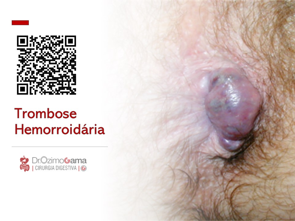
A trombose hemorroidária é uma condição dolorosa e desconfortável que ocorre quando um coágulo de sangue se forma dentro de uma hemorróida. Essa condição é mais comum em gestantes, pessoas com constipação intestinal crônica, e em situações que aumentam a pressão intra-abdominal, como exercícios físicos intensos e levantamento de peso. Neste artigo, exploraremos as causas, sintomas e opções de tratamento para a trombose hemorroidária, ajudando você a entender melhor essa condição e a buscar a melhor abordagem para o seu caso.
Causas da Trombose Hemorroidária
A trombose hemorroidária geralmente está relacionada a fatores de estilo de vida e hábitos pessoais. Os principais gatilhos incluem:
- Obstipação (prisão de ventre): O esforço excessivo para evacuar aumenta a pressão nas veias do ânus, e as fezes endurecidas podem causar traumatismo no tecido anal.
- Gravidez: A pressão adicional no abdômen durante a gravidez pode contribuir para o desenvolvimento da trombose.
- Esforços prolongados e levantamento de peso: Atividades que aumentam a pressão intra-abdominal são fatores de risco.
- Higiene inadequada: A falta de cuidados apropriados na região anal pode exacerbar a condição.
- Fatores adicionais: Permanecer sentado por longos períodos, consumo excessivo de alimentos picantes e bebidas alcoólicas, e prática de sexo anal.
Sintomas da Trombose Hemorroidária
Os principais sinais de trombose hemorroidária incluem:
- Dor intensa na região anal: A dor é geralmente súbita e pode ser bastante severa.
- Sangramento: Frequentemente observado durante a evacuação.
- Inchaço e aumento de volume: Um nódulo na região anal pode se tornar arroxeado ou preto, indicando a presença de um trombo.
Tratamentos Indicados
O tratamento da trombose hemorroidária varia conforme a gravidade da condição. Entre as abordagens recomendadas estão:
- Uso de analgésicos e pomadas anestésicas: Para alívio da dor e desconforto.
- Banhos de assento: Utilizar água morna para aliviar os sintomas e reduzir o inchaço.
- Correção dos hábitos alimentares: Aumentar a ingestão de fibras e líquidos para prevenir a obstipação.
- Tratamento cirúrgico: Em casos mais graves, pode ser necessário realizar uma cirurgia para remover a hemorróida e o trombo.
Se você apresenta sintomas semelhantes, é crucial consultar um médico para uma avaliação adequada e receber o tratamento adequado.
Gostou ❔ Nos deixe um comentário ✍️, ou mande sua dúvida pelo 💬 Chat On-line.
Lembre-se: A informação aqui fornecida é para orientação geral. Sempre consulte seu médico para aconselhamento específico sobre sua situação.
Não Deixe Sua Saúde Esperar! Entre em Contato Conosco Agora!
☎️ CHAT ONLINE 📞
Entre em contato conosco através da nossa central de atendimento dedicada. Nossos especialistas estão prontos para fornecer orientações personalizadas e responder a todas as suas perguntas. Juntos, podemos trabalhar para garantir o melhor para a sua saúde.
🩺(98)991304454 & (98)988664454📲
ATENDIMENTO REMOTO (TELEMEDICINA)
🤳🏻 Agende com os nossos #especialistas pelo WhatsApp ou Telefone
📲 👩🏽⚕️ Clínico: (98) 9 9130 4454
📲 👨🏻⚕️ Cirúrgico: (98) 9 8866 4454
#TromboseHemorroidária #SaúdeDigestiva #TratamentoHemorroidário #BemEstar #CuidadosComASaúde
VEJA AQUI ONDE FAZER UMA AVALIAÇÃO ESPECIALIZADA E RECEBER UMA SEGUNDA OPINIÃO
Leadership in SURGICAL TEAM

Leadership is a process of social influence in which one person can enlist the aid and support of others in the accomplishment of a common task. Successful leaders can predict the future and set the most suitable goals for organizations. Effective leadership among medical professionals is crucial for the efficient performance of a healthcare system. Recently, as a result of various events and reports such as the ‘Bristol Inquiry’, and ‘To Err is Human’ by the Institute of Medicine, the healthcare organizations across different regions have emphasized the need for effective leadership at all levels within clinical and academic fields. Traditionally, leadership in clinical disciplines needed to display excellence in three areas: patient care, research and education.
Within the field of surgery, the last decade has seen various transformations such as technology innovation, changes to training requirements, redistribution of working roles, multi-disciplinary collaboration and financial challenges. Therefore, the current concept of leadership demands to set up agendas in line with the changing healthcare scenario. This entails identifying the needs and initiating changes to allow substantive development and implementation of up-to-date evidence. This article delineates the definition and concept of leadership in surgery. We identify the leadership attributes of surgeons and consider leadership training and assessment. We also consider future challenges and recommendations for the role of leadership in surgery.
PRINCIPLES OF LEADERSHIP FOR GENERAL SURGEONS
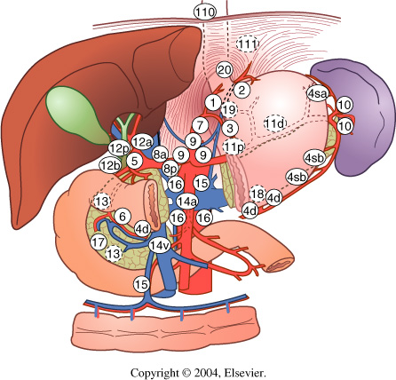
BILIARY-ENTERIC ANASTOMOSIS
 The operative conduct of the biliary-enteric anastomosis centers around three technical steps: 1) identification of healthy bile duct mucosa proximal to the site of obstruction; 2) preparation of a segment of alimentary tract, most often a Roux-en-Y jejunal limb; and 3) construction of a direct mucosa-to-mucosa anastomosis between these two. Selection of the proper anastomosis is dictated by the indication for biliary decompression and the anatomic location of the biliary obstruction. A right subcostal incision with or without an upper midline extension or left subcostal extension provides adequate exposure for construction of the biliary-enteric anastomosis. Use of retractors capable of upward elevation and cephalad retraction of the costal edges are quite valuable for optimizing visual exposure of the relevant hilar anatomy.

Division of the ligamentum teres and mobilization of the falciform ligament off the anterior surface of the liver also facilitate operative exposure; anterocephalad retraction of the ligamentum teres and division of the bridge of tissue overlying the umbilical fissure are critical for optimal visualization of the vascular inflow and biliary drainage of segments II, III, and IV. Cholecystectomy also exposes the cystic plate, which runs in continuity with the hilar plate. Lowering of the hilar plate permits exposure of the left hepatic duct as it courses along the base of segment IVb. In cases of unilateral hepatic atrophy as a result of long-standing biliary obstruction or preoperative portal vein embolization, it is critical to understand that the normal anatomic relationships of the portal structures are altered. In the more common circumstance of right-sided atrophy, the portal and hilar structures are rotated posteriorly and to the right; as a result, the portal vein, which is typically most posterior, is often encountered first; meticulous dissection is necessary to identify the common bile duct and hepatic duct deep within the porta hepatis.
BILIARY-ENTERIC ANASTOMOSIS_THECNICAL ASPECTS
GASTROINTESTINAL STROMAL TUMORS (GIST)

INTRODUÇÃO
GIST, da sigla em inglês gastrointestinal stromal tumors, pertence a um grupo de tumores chamados sarcoma de partes moles. Essa neoplasia se diferencia dos outros tipos de tumores por iniciar-se na parede dos órgãos, junto às camadas musculares do trato gastrointestinal, mais especificamente, nas células do plexo mioenterico, chamadas células de Cajal. Tais células são responsáveis pela motilidade intestinal, sendo consideradas o marca-passo do trato gastrointestinal.
O tumor de GIST é relativamente raro, com estudos atuais mostrando uma prevalência anual em torno de 20 a 40 casos por milhão de habitantes. É mais comum entre pessoas de 50 a 60 anos de idade, sendo extremamente raros até os 20 anos. Por representar um tumor raro, recomenda-se que seja tratado por serviços especializados com cirurgiões do aparelho digestivo, que tenham experiência multidisciplinar na condução e no tratamento dos pacientes com este tumor.
O GIST pode se originar em qualquer local do trato gastrointestinal, do esôfago ao ânus. Em relação à distribuição, 50% a 60% das lesões são provenientes do estômago, 20% a 30% do intestino delgado, 10% do intestino grosso, 5% do esôfago e 5% de outros locais da cavidade abdominal.
DIAGNÓSTICO
A apresentação clínica dos pacientes portadores de GIST não é especifica e depende da localização e do tamanho do tumor. O GIST tem uma característica biológica que é uma mutação genética, com ativação do proto-oncogene Kit e a superexpressão do receptor tirosina quinase (c-Kit). Geralmente, o diagnóstico é feito por uma biópsia da lesão, que a depender da localização, pode ser feita por endoscopia, colonoscopia, ou ecoendoscopia. A tomografia computadorizada do abdômen é importante para avaliação da extensão do tumor e também pode ser utilizada em alguns casos para realização de biópsia do tumor. Não apresentam sinais e/ou sintomas específicos. Podem causar náuseas, vômitos, hemorragias intestinais (vômitos com sangue ou evacuações com sangue ou fezes enegrecidas), sensação de plenitude após alimentação, dor e distensão abdominal, ou presença de uma massa ou tumor palpável no abdômen.
TRATAMENTO
O tratamento padrão para pacientes com GIST não metastático, ou seja, não provenientes de outros órgãos, é a ressecção cirúrgica completa da lesão. Muitas vezes é necessária a cirurgia radical e de grande porte, com a retirada de estruturas e órgãos aderidos, oferecendo a maior chance de cura. O tratamento com imatinib, e mais atualmente ao sunitinib, é utilizado para doença metastática ou irressecável, com intuito de diminuir o tamanho da lesão para que a cirurgia possa ser realizada em melhores condições locais. Tais drogas também podem ser utilizadas após a cirurgia. Para o tratamento sistêmico pode ser necessário estudo genético específico para saber qual a mutação presente no tumor, com intuito de guiar a terapia em relação à dose e tipo de medicação utilizada.
FATORES DE RISCO
Não há fatores de risco diretamente relacionados a essa neoplasia. Manter hábitos de vida saudáveis, uma alimentação balanceada e a prática de exercícios físicos ajudam, de maneira geral, na prevenção do câncer.
PARA MAIS INFORMAÇÕES: http://www.gistsupport.org/
“Not only SURGEONS…”

“… We need a system… which will produce not only surgeons but surgeons of the highest type,…men to study surgery and to devote their energy and their lives to raising the standard of surgical science…”
WS Halsted – Bull Johns Hopkins Hosp 15: 267, 1904.
Avanços no tratamento cirúrgico das METÁSTASES HEPÁTICAS DE ORIGEM COLORRETAL

O câncer colorretal é o terceiro tumor mais frequente no ocidente. Cerca de 50% dos pacientes desenvolvem metástases hepáticas na evolução da doença, as quais são responsáveis por, no mínimo, dois terços das mortes. O avanço nas técnicas cirúrgicas e a melhora dos esquemas quimioterápicos têm permitido oferecer tratamento com intuito curativo a um número cada vez maior de pacientes. Os avanços recentes do tratamento das metástases hepáticas, incluindo estratégias para aumentar as ressecções (por exemplo: embolização da veia porta, ablação por radiofrequência, hepatectomia em dois tempos, quimioterapia de conversão e estratégia inversa de tratamento) e hepatectomias na presença de doença extra-hepática possibilitam uma melhor sobrevida dos pacientes.
TRANSPLANTE DE FÍGADO
Este artigo é destinado a pacientes que irão passar por um transplante hepático e seus familiares. Ele visa esclarecer dúvidas comuns e fornecer informações importantes para enfrentar a experiência de forma mais tranquila e informada.
1. Quando está indicado o transplante?
O transplante hepático é recomendado em casos de:
- Cirrose Hepática: Quando exames mostram função hepática diminuída ou sintomas como ascite, sangramento digestivo, encefalopatia hepática ou peritonite bacteriana.
- Hepatocarcinoma: Tumores originários do fígado, muitas vezes associados à cirrose, podem ser tratados com transplante.
- Colangite Esclerosante e Obstrução Intratável de Vias Biliares: Doenças que causam obstrução das vias biliares e infecções graves.
- Hepatite Fulminante e Perda do Fígado Transplantado: Em casos de hepatite grave ou perda do enxerto transplantado dentro de 30 dias, a urgência é priorizada.
2. Quais são as contra-indicações ao transplante hepático?
Não é indicado realizar o transplante em situações como:
- Doença avançada em outros órgãos.
- Tumores fora do fígado.
- Abuso atual de drogas ou álcool.
- Incapacidade de seguir orientações médicas.
- Infecção ativa (o transplante pode ser realizado após a infecção ser controlada).
A idade avançada não é mais uma contra-indicação absoluta, mas deve ser avaliada individualmente.
3. A fila de transplante
Atualmente, a fila de transplante no Brasil é organizada pelo sistema Meld/Peld, que prioriza pacientes com base na gravidade da doença. A fila é subdividida por grupo sanguíneo (A, B, AB, O) e compatibilidade com o tamanho do paciente. Pacientes com grupo sanguíneo raro (AB) podem receber órgãos de todos os grupos.
4. O fígado “não ideal” (marginal)
Órgãos “não ideais” ou marginais podem ter características menos perfeitas, como mais gordura ou doadores com condições subótimas. Eles são destinados a pacientes com necessidades urgentes, como aqueles com tumores hepáticos.
5. A equipe
A equipe de transplante é composta por diversos profissionais, incluindo médicos, enfermeiros, nutricionistas, psicólogos e fisioterapeutas. Cada paciente passa por avaliações detalhadas de todos esses profissionais.
6. Como proceder em caso de emergências durante a espera pelo transplante
Em situações urgentes, dirija-se ao pronto-socorro ou entre em contato com sua equipe médica. As emergências incluem:
- Vômitos com sangue.
- Fezes com sangue ou escurecidas.
- Queda de pressão, mal-estar súbito.
- Colangite (febre, dor abdominal, icterícia).
- Peritonite (infecção abdominal).
7. As reuniões com a Coordenação de Enfermagem de Transplantes
Reuniões regulares são realizadas para pacientes na fila e seus familiares, oferecendo a oportunidade de trocar experiências, fazer perguntas e receber atualizações sobre a lista de espera.
8. A doação de órgãos através de doador em morte encefálica
A doação é um ato voluntário feito com consentimento familiar. O paciente deve estar em morte encefálica, uma condição onde não há atividade cerebral, mas o coração e outros órgãos ainda funcionam. A entidade responsável é o Rio Transplante e os dados do doador são confidenciais.
9. Chamada para o transplante
Mantenha-se disponível e acessível para a equipe de transplante, com telefone atualizado e a uma distância máxima de duas horas do hospital. Caso seja chamado, esteja preparado para o possível ajuste no órgão doado.
A CIRURGIA DE TRANSPLANTE HEPÁTICO
A cirurgia dura entre 5 a 8 horas e é dividida em duas etapas principais:
- Captação: Retirada do fígado doado, que pode ser considerado apto, marginal ou inapto.
- Transplante: O fígado doente é retirado e o novo órgão é implantado, com a realização das anastomoses das veias, artéria hepática e via biliar.
Pós-operatório Imediato
Você será monitorado na UTI por aproximadamente 48 horas. Equipamentos como tubos respiratórios, eletrodos, sondas e drenos serão usados para garantir segurança e monitoramento.
Pós-operatório
A internação média é de 2 semanas, variando conforme o paciente. É importante realizar exames regularmente e seguir orientações médicas para movimentação e exercícios respiratórios.
Medicação
Os imunosupressores, fornecidos pelo SUS, são essenciais para evitar a rejeição do transplante. Familiarize-se com o processo de obtenção e renovação desses medicamentos. Outras medicações de rotina devem ser providenciadas pela família.
Intercorrências após o Transplante
Monitorar sinais de rejeição e infecções é crucial. Mantenha-se saudável, evite álcool e siga as orientações médicas rigorosamente.
Alta Hospitalar
Antes da alta, você receberá orientações detalhadas e agendamentos para consultas e exames. Siga as recomendações de fisioterapia, nutrição e outros cuidados necessários.
Seguimento Inicial e Tardio
O acompanhamento pós-transplante é fundamental. As consultas variam ao longo do tempo, com exames laboratoriais e visitas regulares ao hospital.
Efeitos Colaterais
Os imunosupressores podem causar efeitos colaterais, como tremores e aumento da glicose. Informe seu médico sobre qualquer efeito adverso e não suspenda a medicação por conta própria.
Cuidados Gerais
Controle o peso, colesterol, glicose e pressão arterial. Evite bebidas alcoólicas, use máscara em locais públicos, evite contato com pessoas doentes e exposição solar excessiva. Mantenha acompanhamento ginecológico e dentário regular e consulte seu médico antes de tomar novos medicamentos. Este artigo é uma referência para ajudar você a entender e gerenciar o processo de transplante hepático. Mantenha-se em contato com sua equipe médica para qualquer dúvida ou preocupação.
PRESCRIÇÃO MÉDICO-CIRÚRGICA

“de nada adianta um diagnóstico brilhante se o seu tratamento não for compreendido.”
A cirurgia é uma forma de terapia para as doenças que é motivada pelo CORAÇÃO, planejada na CABEÇA e executada pelas MÃOS.
“A cirurgia é uma forma de terapia para as doenças que é motivada pelo CORAÇÃO, planejada na CABEÇA e executada pelas MÃOS.”
Prof . Phd. Tom DeMeester
DEFINIÇÃO DE CIRURGIA (By Tom DeMeester / Gastrão 2012)
Lesão de Vias Biliares na Colecistectomia: Prevenção e Tratamento
A via laparoscópica tem sido reconhecida como padrão de excelência para a colecistectomias. Phillipe Mouret foi quem primeiro a realizou em 1987, mas outros procedimentos já haviam sido realizados por laparoscopia e foram descritos por ginecologistas. Desenvolvida no final da década de 80 e início dos anos 90, a videolaparoscopia mudou os conceitos de acesso cirúrgico e campo operatório, introduzindo a concepção de “cirurgia minimamente invasiva”.A colecistectomia é um dos procedimentos cirúrgicos mais realizados no mundo. Com o advento da videolaparoscopia, tornou-se uma cirurgia menos traumática, mais estética, com períodos mais curtos de internação. Em contrapartida, observou-se o aumento da incidência de lesões de via biliar extra-hepática quando comparado ao procedimento aberto, fato preocupante devido à morbidade elevada desse tipo de lesão, cuja mortalidade não é desprezível.
VALORES ÉTICOS DOS ESTAGIÁRIOS DA CLÍNICA CIRÚRGICA
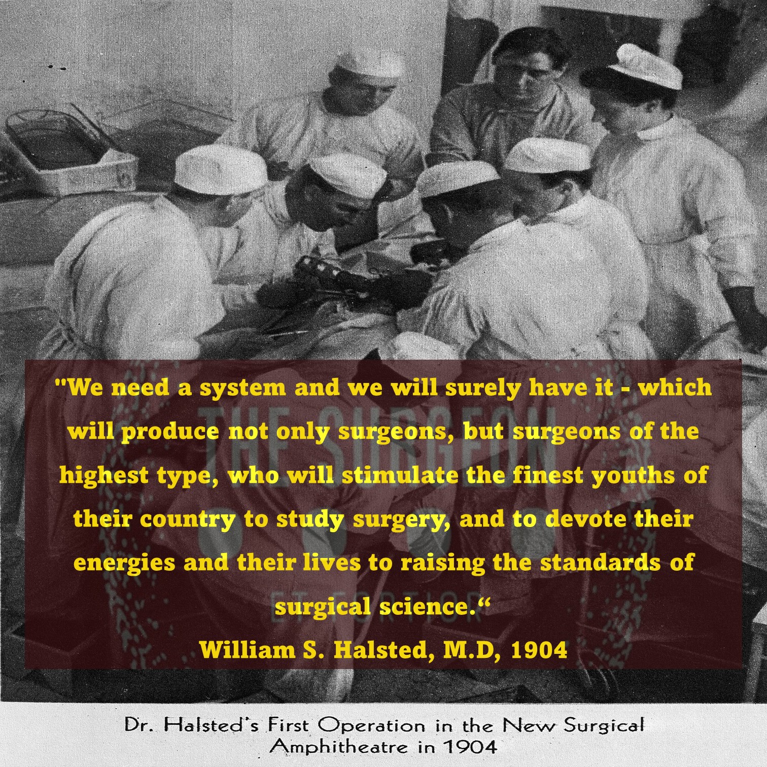
EXCELÊNCIA
Nunca baixar os seus padrões pessoais e profissionais. Fazer sempre o melhor, NÃO SOMENTE O POSSÍVEL.
Fazer sempre a coisa certa, mesmo quando ninguém está olhando.
RESPONSABILIDADE
Cumprir rigorosamente as atribuições em relação aos cuidados com os pacientes do serviço.
Ser uma força positiva com comprometimento na melhor assistência ao paciente.
DISCIPLINA
É a construção diária do seu objetivo.
O sucesso vem um pequeno passo de cada vez. O amanhã começa agora.

Surgical Rotation: 5 PRINCIPLES

1.Getting along with the nurses.
The nurses do know more than the rest of us about the codes, routines, and rituals of making the wards run smoothly. They may not know as much about pheochromocytomas and intermediate filaments, but about the stuff that matters, they know a lot. Acknowledge that, and they will take you under their wings and teach you a ton!
2. Helping out.
If your residents look busy, they probably are. So, if you ask how you can help and they are too busy even to answer, asking again probably would not yield much. Always leap at the opportunity to shag x-rays, track down lab results, and retrieve a bag of blood from the bank. The team will recognize your enthusiasm and reward your contributions.
3. Getting scutted.
We all would like a secretary, but one is not going to be provided on this rotation. Your residents do a lot of their own scut work without you even knowing about it. So if you feel like scut work is beneath you, perhaps you should think about another profession.
4. Working hard.
This rotation is an apprenticeship. If you work hard, you will get a realistic idea of what it means to be a resident (and even a practicing doctor) in this specialty. (This has big advantages when you are selecting a type of internship. Staying in the loop. In the beginning, you may feel like you are not a real part of the team. If you are persistent and reliable, however, soon your residents will trust you with more important jobs. Educating yourself, and then educating your patients. Here is one of the rewarding places (as indicated in question 1) where you can soar to the top of the team. Talk to your patients about everything (including their disease and therapy), and they will love you for it.
5. Maintaining a positive attitude.
As a medical student, you may feel that you are not a crucial part of the team. Even if you are incredibly smart, you are unlikely to be making the crucial management decisions. So what does that leave: attitude. If you are enthusiastic and interested, your residents will enjoy having you around, and they will work to keep you involved and satisfied. A dazzlingly intelligent but morose complainer is better suited for a rotation in the morgue. Remember, your resident is likely following 15 sick patients, gets paid less than $2 an hour, and hasn’t slept more than 5 hours in the last 3 days. Simple things such as smiling and saying thank you (when someone teaches you) go an incredibly long way and are rewarded on all clinical rotations with experience and good grades.
Having fun! This is the most exciting, gratifying, rewarding, and fun profession and is light years better than whatever is second best (this is not just our opinion).
By: Alden H. Harken MDProfessor and Chair, Department of Surgery, University of California, San Francisco–East Bay, Oakland, California, Chief of Surgery, Department of Surgery, Alameda County Medical Center, Oakland, California
AVALIAÇÃO PRÉ-OPERATÓRIA
MEDICINA BASEADA EM EVIDÊNCIA_AVALIAÇÃO PRÉ_OPERATÓRIA

O médico CIRURGIÃO realiza a avaliação pré-operatória e define a necessidade de avaliação complementar, considerando a otimização das condições clínicas do paciente e a realização de exames complementares. Pacientes hígidos, com idade inferior a 40 anos, sem fatores de risco detectados na anamnese e no exame físico, a serem submetidos a cirurgias de pequeno porte, após a avaliação clínica básica poderão ser encaminhados à cirurgia após avaliação do Anestesiologista. Porém, nos casos em que o paciente tenha alguma comorbidade, idade maior de 60 anos, estado físico ASA II ou acima, obesidade, dependência funcional ou cirurgia de médio ou grande porte, estará indicado avaliação pré-operatória mais pormenorizada .
“poderosos além de qualquer medida…”

Nosso medo mais profundo não é o de sermos inadequados. Nosso medo mais profundo é que somos poderosos além de qualquer medida. É a nossa luz, não nossa escuridão, que mais nos assusta. Nós nos perguntamos: Quem sou eu para ser brilhante, maravilhoso, talentoso e fabuloso? Na verdade, quem não quer que você seja? Você é um filho de Deus. Seu papel pequeno não serve ao mundo. Não há nada de iluminado em se encolher, para que outras pessoas não se sintam inseguros ao seu redor. Estamos todos feitos para brilhar, como as crianças. Nascemos para manifestar a glória de Deus que está dentro de nós. Não é apenas em alguns de nós, está em todos. E conforme deixamos nossa própria luz brilhar, inconscientemente damos às outras pessoas permissão para fazer o mesmo. Como estamos libertamos do nosso medo, nossa presença, automaticamente, libera os outros.
Esse texto é atribuído a Marianne Williamson, uma autora e palestrante americana. A passagem faz parte de seu livro “A Return to Love: Reflections on the Principles of A Course in Miracles” (1992). Williamson explora temas de autoaceitação, potencial humano e espiritualidade, incentivando as pessoas a reconhecerem e expressarem sua luz interior. A citação destaca a ideia de que o verdadeiro medo não está em nossa inadequação, mas no poder e potencial que temos para impactar positivamente o mundo ao nosso redor.
Eventos Adversos na Clínica Cirúrgica
EVENTOS ADVERSOS # COMPLICAÇÕES

Introdução
O termo “Evento Adverso (EA)” cirúrgico é relativamente novo, mas o conceito de monitoramento dos resultados cirúrgicos, incluindo complicações pós-operatórias, é muito antigo. Desde 1732, existem referências a sistemas de coleta de informações hospitalares. Na Grã-Bretanha, estatísticas vitais datam de 1838, e já em 1850, a associação entre higiene das mãos e a transmissão de infecções foi estabelecida. Em 1854, os riscos de má higiene nos hospitais foram destacados, e em 1910, Ernest Codman defendeu a avaliação rotineira dos resultados negativos em cirurgias para melhorar a qualidade da assistência. Nos anos 1990, o interesse em erros e danos relacionados à saúde cresceu, mudando o foco das pesquisas para estratégias de enfrentamento e uma abordagem sistêmica ou organizacional.
Epidemiologia
Os EAs cirúrgicos contribuem significativamente para a morbidade pós-operatória, e sua avaliação e monitoramento frequentemente são imprecisos. Com a redução do tempo de permanência hospitalar e o aumento do uso de técnicas cirúrgicas inovadoras, especialmente minimamente invasivas e endoscópicas, a eficiência no monitoramento dos eventos adversos torna-se crucial. Revisões recentes identificaram que os EAs são desfavoráveis, indesejáveis, prejudiciais, impactam o paciente e estão associados ao processo de assistência à saúde, mais do que a um processo natural de doenças. A análise dos EAs é complexa devido à variabilidade dos sistemas de registro e às diversas definições na literatura científica para complicações pós-operatórias.
Custo e Efeitos de Medicina Legal
As complicações pós-operatórias resultam da interação de fatores dependentes do paciente, sua enfermidade e a atenção à saúde recebida. O estudo dos EAs cirúrgicos é relevante pela sua frequência, impacto considerável sobre a saúde dos pacientes e repercussão econômica no gasto social e sanitário. Além disso, os EAs servem como instrumento de avaliação da qualidade da assistência. Eventos adversos evitáveis, suscetíveis a intervenções de prevenção, são de maior interesse para a saúde pública. EAs cirúrgicos estão relacionados a acidentes intra-operatórios, complicações pós-operatórias imediatas ou tardias e ao fracasso da intervenção cirúrgica.
Nos Estados Unidos, um estudo em hospitais de Colorado e Utah calculou uma taxa de incidência de 1,9% para pacientes internados, e 3,0% para pacientes submetidos a cirurgia ou parto, com 54% dos EAs considerados evitáveis. Em 5,6% dos casos, os EAs resultaram em óbito. Na Austrália, a prevalência de internações cirúrgicas associadas a um EA foi de 21,9%, com 47,6% dos EAs classificados como altamente evitáveis. Na Espanha, um estudo em cirurgias de parede abdominal encontrou complicações em 16,32% dos pacientes.
Diferença entre Complicação e Evento Adverso
Complicações pós-operatórias surgem da interação entre fatores do paciente, da doença e da atenção recebida. Eventos Adversos, por outro lado, são desfavoráveis, indesejáveis e prejudiciais, resultantes do processo de assistência à saúde. Embora ambos afetem a recuperação, os EAs são frequentemente evitáveis com intervenções preventivas.
Conclusão
A segurança em cirurgia no Brasil é preocupante. Em 2003, 52,5% dos hospitais inspecionados pelo Conselho Regional de Medicina de São Paulo apresentaram condições físicas inadequadas. Hospitais de pequeno porte, que representam 62% dos estabelecimentos, enfrentam desafios significativos em termos de complexidade e densidade tecnológica.
Como Ernest Codman sabiamente afirmou: “A melhoria na assistência médica só pode ser alcançada com a análise sistemática dos resultados”. Esta frase histórica ressoa ainda hoje, destacando a importância da avaliação e monitoramento dos EAs cirúrgicos para a melhoria contínua da qualidade e segurança na assistência à saúde.
Tratamento Cirúrgico do HEMANGIOMA HEPÁTICO

Introdução
O hemangioma é o tumor hepático mais comum, identificado em 5% a 7% das necropsias. Sua incidência é maior entre a terceira e a quinta décadas de vida, com prevalência nas mulheres. Este tumor pode aumentar de tamanho durante a gravidez e com o uso de estrogênios.
Conceito
Hemangiomas hepáticos são tumores benignos dos vasos sanguíneos do fígado. Apesar de sua causa não estar completamente esclarecida, há indícios do papel dos hormônios sexuais em seu desenvolvimento. Observações como a presença de receptores de estrogênio em alguns hemangiomas e o aumento de tamanho durante a puberdade, gravidez e uso de anticoncepcionais sugerem essa relação.
Epidemiologia
A maioria dos hemangiomas hepáticos é única e mede menos de 4 cm de diâmetro, com apenas 10% sendo múltiplos e podendo alcançar até 27 cm. Hemangiomas gigantes são definidos como aqueles com mais de 4 cm de diâmetro. Em geral, o tamanho da maioria dos hemangiomas permanece inalterado ao longo do tempo.
Diagnóstico
O diagnóstico de hemangioma hepático é geralmente realizado por exames de imagem, como tomografia computadorizada e ressonância magnética. Esses exames identificam padrões típicos de impregnação nodular periférica e descontínua, com aumento gradual da impregnação. Na ressonância magnética, os hemangiomas tipicamente apresentam um sinal alto nas sequências ponderadas em T2. A cintilografia com hemácias marcadas também é precisa para hemangiomas maiores que 2 cm, mas raramente necessária.
Indicações de Tratamento Cirúrgico
A maioria dos hemangiomas não requer tratamento. Não há evidências para interromper o uso de anticoncepcionais hormonais ou evitar a gravidez em pacientes com hemangioma, mesmo em casos de tumores gigantes. Complicações são raras, mas incluem inflamação, coagulopatia, sangramento e compressão de estruturas vizinhas. O tratamento cirúrgico é indicado apenas em casos de sintomas significativos, crescimento tumoral, síndrome de Kasabach-Merritt, ou quando não é possível excluir malignidade.
Conclusões
Hemangiomas hepáticos, em sua maioria, são assintomáticos e não necessitam de intervenção. O tratamento cirúrgico, apesar de raramente necessário, é realizado em casos selecionados para evitar complicações maiores. A decisão cirúrgica deve ser cuidadosa, considerando os riscos operatórios. Como disse William Osler, “A prática da medicina é uma arte, não um comércio; uma vocação, não um negócio; uma chamada em que seu coração será exercitado igualmente com sua cabeça.”
World’s Greatest Surgeon
 On July 12, 2008, the world lost an incredible talent. A renegade physician, a pioneer, the father of open-heart surgery, and perhaps the best surgeon who ever lived, Dr. Michael DeBakey died of natural causes at 99. Because of his groundbreaking research, cutting-edge medical devices and maverick approach to cardiac surgery, DeBakey literally changed the rules of the game and thousands of lives are saved each day.
What can we learn from Michael DeBakey’s life and career?
1. Build your brand.
With a career that spanned more than 70 years, DeBakey built a reputation for being indispensable. His patients included everyone from the ordinary person next door and people with no means to a list of Who’s Who among world leaders. Presidents Kennedy, Johnson and Nixon, President Boris Yeltsin, King Hussein of Jordan, the Shah of Iran, Turkish President Turgut Ozal, just to name a few, engaged DeBakey because they knew he was the best. The Journal of the American Medical Association said in 2005, “Many consider Michael E. DeBakey to be the greatest surgeon ever.” Is your personal brand strong enough that if you left your company, colleagues and customers would have a difficult time getting along without you?
2. Be a guru, thought leader, industry expert.
Dr. DeBakey published more than 1,000 medical reports, research papers, chapters and books on topics related to cardiovascular medicine. He helped establish the National Library of Medicine, the world’s largest and most prestigious repository of medical archives. DeBakey played a key role in organizing a specialized medical center system to treat soldiers returning from the war. This system is now the Veterans’ Administration Medical Center System. For his numerous contributions Dr. DeBakey was awarded the Presidential Medal of Freedom, the Congressional Gold Medal, Congress’ highest civilian honor, the National Medal of Science, the country’s highest scientific award, and The United Nations Lifetime Achievement Award. Do people see you as a guru in your field? How distinctive is your knowledge base? How well do you garner, contribute and leverage knowledge?
3. Never quit learning.
As a child, DeBakey was required to borrow a book from the library each week and read it. He read the entire Encyclopedia Britannica before entering high school. Overseeing cases, consulting with colleagues and mentoring younger surgeons, he made his mark on the world right up to the end. DeBakey performed his last surgery at age 90 and continued to travel the globe giving lectures. Perhaps you’re thinking, “Who would want a 90-year-old surgeon operating on them?” The answer could be, “Someone who’s performed more than 60,000 cardiovascular procedures in his career.” Do you have a reputation for lifelong learning, for continually adding value? When we stop bringing something new to the game, the game is over.
4. Risk more, gain more.
DeBakey took risks others weren’t willing to take to advance medicine. Tubing, clamps, pumps, protocols all bear the mark of DeBakey’s passion for innovation. Yet, product and process innovations often pull people out of their comfort zones and some of DeBakey’s early breakthroughs weren’t accepted initially—in fact they were ridiculed. For example, in 1939, when Drs. DeBakey and Alton Ochsner linked cigarette smoking to lung cancer, many in the medical community derided it. Then in 1964, the Surgeon General confirmed their findings and documented the cause and effect. There was also skepticism when DeBakey discovered that he could substitute parts of diseased arteries with synthetic (Dacron) grafts—a procedure that enables surgeons to repair aortic aneurysms in the chest and abdomen. He initially figured out how to stitch synthetic blood vessels on his wife’s sewing machine. Now the procedure is widely used. DeBakey was also the first to perform bypass surgery and the first to perform a successful removal of a blockage of the carotid (main) artery of the neck, a procedure that has become the standard protocol for treating stroke. The world is not changed by those who are unwilling to take risks. Is your passion for advancing your field by taking a risk bigger than your fear of rejection or making a mistake?
5. Refuse to sell out on your dream.
DeBakey developed an interest in medicine in his father’s pharmacy where he listened to physicians talk shop. The vision to become a doctor was clear, the question was, “what kind?” In 1932, there simply wasn’t anything you could do for heart disease, if a patient had a heart attack the long-term prognosis wasn’t good. While he was still in school in 1932, DeBakey invented the roller pump—a critical part of the heart-lung machine that takes over the functions of the heart and lungs during open-heart surgery. This not only created the era of open-heart surgery, it cemented DeBakey’s passion to make a mark in the world of cardiovascular medicine. Engagement is about pouring your heart, mind and soul into a dream that causes you to fire on all cylinders. Does your career fulfill your desires? Or, have you sacrificed a dream that could make you come alive for a life of duty and routine that simply “works”?
6. Play to your genius.
DeBakey said, “I like my work, very much. I like it so much that I don’t want to do anything else.” Most people who are happy in life spend time doing what they love. This usually makes them extremely good at what they do. Dr. DeBakey exemplified the power of what can happen when our work requires what we are good at and passionate about. Playing to your genius is about using your gifts and talents to pursue a passion that makes a significant contribution to the people and the world you serve. Playing to your genius also promotes autonomy and self-direction, cultivates commitment, stimulates personal growth and makes work fun. Are you engaged in work you’re good at and passionate about—work that makes a contribution and needs to be done? Or are you just biding time?
7. Balance passion with discipline and focus.
With regard to his patients, the indefatigable DeBakey had an uncompromising dedication to perfection. He was known as a taskmaster who set very high standards, yet he never demanded more from others than he demanded from himself. Heart surgeons who trained under DeBakey say he was hard to keep up with when making patient rounds. They joked that he was from another world because he could maintain his focus and intensity for hours. In a world of competing priorities and information overload it’s easy to lose focus and get distracted. But, if you are playing to your genius and doing what you love, it’s easier to be disciplined and maintain a maniacal focus. Are you disciplined? Do you have a maniacal focus? Would your customers (internal and external) say you are relentless when it comes to pursuing perfection?
8. Find a void and figure out how to fill it.
Michael DeBakey’s innovations are on par with the likes of Thomas Edison, Alexander Graham Bell, Jonas Salk, Henry Ford and Alfred Nobel. During World War II, he helped establish the mobile army surgical hospitals or MASH units. He was a key player in the development of artificial hearts, artificial arteries and bypass pumps that help keep patients alive who are waiting for transplants. He was among the first to recognize the importance of blood banks and transfusions. He also helped create more than 70 surgical instruments that made procedures easier and clinical outcomes more effective. If something couldn’t be done, DeBakey found a way to do it. In 1967, Dr. Christiaan Barnard performed the first human heart transplant in South Africa. Dr. DeBakey was among the first to begin doing the procedure in the United States. The problem was that recipients’ bodies rejected the new organs and death rates were high. In the 1980s cyclosporine, a new anti-rejection drug paved the way for organ transplants. Again, DeBakey was among the first to develop new protocols and advance the field of heart transplants. Where are the gaps in your organization or industry? What would happen if you developed a reputation for filling these voids?
9. Show people that their work matters.
Michael DeBakey is known not only for his prolific contributions to the medical field, but also as a symbol of hope and encouragement to his colleagues. Many years ago a colleague of ours shadowed Dr. DeBakey for a day at The Methodist Hospital in Houston, Texas. He was struck by DeBakey’s capacity to affirm each person he saw in the course of the day. In one particular encounter, DeBakey began chatting with an elderly janitor who was sweeping the floor. DeBakey asked the man about his wife and children. He told the older man, obviously not for the first time, that the hospital couldn’t function without the janitor because germs would spread, increasing the chances of infection in the hospital. Later in the day, our colleague tracked down the janitor and asked him, “What exactly do you do? Tell me about your job.” With pride, the janitor replied: “Dr. DeBakey and I? We save lives together.” He’s right. After all, consider what would happen to our healthcare systems if the cleaning crews went on strike. DeBakey understood that showing the janitor exactly how he contributes to a larger, more heroic cause is crucial. This creates a powerful dynamic. Realizing that he is working toward a worthy goal, the janitor’s perceptions about his work changed. It had new meaning and his enthusiasm for the job was rejuvenated. Great leaders make time to help people see how their work is connected to something bigger. For a surgeon like DeBakey, those five or ten minutes each day were costly, unless, of course, you consider the productivity generated by a janitor whose work has been transformed. Right now, how many people in your organization are engaged in work that five years from today no one will give a rip about? Can you make the link between what you do and a noble or heroic cause? Can you make this link for others?
10. Be generative—inspire others to pursue the cause.
Generativity is the care and concern for the development of future generations through teaching, mentoring, and other creative contributions. It’s about leaving a positive legacy. All great leaders are generative and Michael DeBakey was no exception. He inspired many medical students to pursue careers in cardiovascular surgery. His reputation brought many people to Baylor College of Medicine and helped transform it into one of the premier medical institutions in the world. DeBakey trained and mentored almost 1,000 surgeons and physicians. In 1976, his students founded the Michael E. DeBakey International Surgical Society. Many of his residents went on to serve as chairpersons and directors of their own successful academic surgical programs in the United States and around the world. Are the people you’ve touched in your career learning, growing and making a difference as a result of your influence? Have they been inspired to build a better world than the world they inherited? Michael DeBakey applied his problem-solving skills to many parts of medicine that have changed our way of life. Timothy Gardner, M.D., president of the American Heart Association said it well, “DeBakey’s legacy will live on in so many ways—through the thousands of patients he treated directly and through his creation of a generation of physician educators, who will carry his legacy far into the future. His advances will continue to be the building blocks for new treatments and surgical procedures for years to come.”
Michael DeBakey’s life and legacy proves that one person who chooses to play to their genius can change the world and make it a better place for all. What legacy will you leave behind?
O CIRURGIÃO (POEMA)
O CIRURGIÃO
Um corpo inerte aguarda sobre a mesa
Naquele palco despido de alegria.
O artista das obras efêmeras se apresenta.
A opereta começa, ausente de melodia
E o mascarado mudo trabalha com presteza.
Sempre começa com esperança e só términa com certeza.
Se uma vida prolonga, a outra vai-se escapando.
E nem sempre do mundo o aplauso conquistando
Assim segue o artista da obra traiçoeira e conquistas passageiras.
Há muito não espera do mundo os louros da vitória
Estudar, trabalhar é sua história, e a tua maior glória
Hás de encontrar na paz do dever cumprido.
Quando a vivência teus cabelos prateando
E o teu sábio bisturi, num canto repousando
Uma vez que sua missão vai terminando
Espera do bom Deus por tudo, a ti, seja piedoso.
SOIS VÓS INSTRUMENTO DA TUA OBRA.
.
The Qualities of a GOOD SURGEON

Following is a list of Dr. Ephraim McDowell’s personal qualities described as “C” words along with evidence corroborating each of the characteristics.
Courageous: When he agreed to attempt an operation that his teachers had stated was doomed to result in death, he, as well as his patient, showed great courage.
Compassionate: He was concerned for his patient and responded to Mrs. Crawford’s pleas for help.
Communicative: He explained to his patient the details of her condition and her chances of survival so that she could make an informed choice.
Committed: He promised his patient that if she traveled to Danville, he would do the operation. He made a commitment to her care.
Confident: He assured the patient that he would do his best, and she expressed confidence in him by traveling 60 miles by horseback to his home.
Competent: Although lacking a formal medical degree, he had served an apprenticeship in medicine for 2 years in Staunton, Virginia, and he had spent 2 years in the study of medicine at the University of Edinburgh, an excellent medical school. In addition, he had taken private lessons from John Bell, one of the best surgeons in Europe. By 1809 he was an experienced surgeon.
Carefull: Despite the fact that 2 physicians had pronounced Mrs. Crawford as pregnant, he did a careful physical examination and diagnosed that she was not pregnant but had an ovarian tumor. He also carefully planned each operative procedure with a review of the pertinent anatomic details. As a devout Presbyterian, he wrote special prayers for especially difficult cases and performed many of these operations on Sundays.
Courteous: He was humble and courteous in his dealings with others. Even when he was publicly and privately criticized after the publication of his case reports, he did not react with vitriol. The qualities of character demonstrated by Dr. Ephraim McDowell 200 years ago are still essential for surgeons today.
Dr. Ephraim McDowell exemplified the essential qualities that define a great surgeon. His courage was evident when he accepted to perform an operation deemed fatal by his mentors, facing the unknown with determination. His compassion shone through in his genuine concern for the well-being of his patients, responding to cries for help with sensitivity and empathy. He was communicative, ensuring that his patients were well-informed about their conditions and the risks involved, enabling decisions based on knowledge. Additionally, Dr. McDowell demonstrated unwavering commitment to his patients, keeping promises and ensuring that each received the best possible care.
His confidence in his abilities, even without a formal degree, was supported by a robust education and a continuous dedication to learning. Dr. McDowell was competent, the result of years of study and practice, conducting meticulous physical examinations and planning each procedure with precision. His courtesy and humility in interactions, even in the face of criticism, showed a character that valued respect and integrity. These qualities, demonstrated two centuries ago, are timeless and continue to define ethical practice in modern surgery. As William Osler aptly stated, “The practice of medicine is an art, not a trade; a calling, not a business; a calling in which your heart will be exercised equally with your head.”
Tratamento Cirúrgico da COLECISTITE AGUDA
I. INTRODUÇÃO
Os cálculos de vesícula (COLELITÍASE) estão presentes em mais de 10% da população ocidental e esta incidência aumenta com a idade. A colelitíase é a doença do aparelho digestivo com maior número de indicação cirúrgica. Anualmente, cerca de 200.000 colecistectomias são realizadas nos Brasil. Os fatores de risco para o surgimento dos cálculos são: obesidade, diabetes mellitus, uso de estrogênio, gravidez, doença hemolítica, hereditariedade e cirrose. Acomete principalmente as mulheres na idade reprodutiva.
São várias as complicações da colelitíase, entre elas:
1. COLECISTITE AGUDA
2. PANCREATITE AGUDA
3. COLEDOCOLITÍASE
4. FÍSTULAS INTERNAS
5. CÂNCER DA VESÍCULA
VEJA AQUI ONDE REALIZAR A CIRURGIA DE COLECISTECTOMIA COM CIRURGIÃO DO APARELHO DIGESTIVO
II. FISIOPATOLOGIA
A colecistite aguda é uma doença comum em emergências em todo o mundo. Na maioria dos casos, é causada pela inflamação da parede da vesícula secundária à impactação de um cálculo no ducto cístico obstruindo-o, o que causa uma crise repentina de dor abdominal, conhecido como ABDOME AGUDO.
Colecistite litiásica
A colecistite aguda está associada à colelitíase em mais de 90% dos casos. O quadro ocorre devido à obstrução do ducto cístico por um cálculo. Se a obstrução continua, a vesícula se distende e suas paredes tornam-se edematosas. O processo inflamatório inicia-se com espessamento da parede, eritema e hemorragia subserosa. Surgem hiperemia e áreas focais de necrose. Na maioria dos casos, o cálculo se desloca e o processo inflamatório regride. Se o cálculo não se move, o quadro evolui para isquemia e necrose da parede da vesícula em cerca de 10% dos casos. A formação de abscesso e empiema dentro da vesícula é conhecida como colecistite aguda gangrenosa. Com a infecção bacteriana secundária, principalmente por anaeróbios, há formação de gás que pode ocorrer dentro ou na parede da vesícula. Esse é um quadro mais grave conhecido com colecistite enfisematosa.
Colecistite aguda alitiásica
A colecistite aguda também pode ocorrer sem a presença de cálculos em cerca de 5% dos casos. Tem uma evolução mais rápida e frequentemente evolui para gangrena, empiema ou perfuração. Ocorre em pessoas idosas ou em estado crítico após trauma, queimaduras, nutrição parenteral de longa data, cirurgias extensas, sepses, ventilação com pressão positiva e a terapia com opioides também parece estar envolvida. A etiologia é confusa, mas a estase, a isquemia, a injúria por reperfusão e os efeitos dos mediadores pró-inflamatórios eicosanoides são apontados como causas.
VEJA AQUI ONDE REALIZAR A CIRURGIA DE COLECISTECTOMIA COM CIRURGIÃO DO APARELHO DIGESTIVO
III. QUADRO CLÍNICO
O quadro se inicia com uma cólica biliar caracterizada como dor no hipocôndrio direito com irradiação para escápula direita e região epigástrica. Como sintoma mais comum, o paciente apresenta dor e pressão no hipocôndrio direito, mais duradoura das que nas crises de cólica biliar a que ele frequentemente se refere. Esse é o primeiro sinal de inflamação da vesícula. A dor pode intensificar-se quando a pessoa respira profundamente e muitas vezes estende-se à parte inferior da escápula direita e à região epigástrica. A febre, assim como náuseas e vômitos, que podem ser biliosos, são habituais em 70% dos pacientes. A febre alta, os calafrios e a distensão abdominal com diminuição da peristalse costumam indicar a formação de um abscesso, gangrena ou perfuração da vesícula biliar. Nestas condições, torna-se necessária a cirurgia de urgência. A icterícia pode indicar coledocolitíase ou compressão externa do colédoco pela vesícula inflamada.
IV. AVALIAÇÃO DIAGNÓSTICA
O hemograma habitualmente apresenta leucocitose com desvio para esquerda. O hepatograma está alterado com elevação das transaminases, da fosfatase alcalina, bilirrubinas e amilase. A hiperbilirrubinemia pode ser devido à compressão extrínseca pelo processo inflamatório grave, pela coledocolitíase ou pela síndrome de Mirizzi, que é causada pela impactação de um cálculo no infundíbulo que pode fistulizar para o colédoco e obstruí-lo. A hiperamilasemia pode ocorrer devido à obstrução do ducto pancreático levando à pancreatite concomitante.
A ultrassonografia é o exame inicial e permite a identificação de alterações que não são visíveis no exame físico e permite uma classificação. É considerado o exame “ouro” nesses casos. Tem alta sensibilidade para a detecção de cálculos e o espessamento da parede que é considerado anormal quando maior que 4 mm . Também pode haver visualização de líquido perivesicular, distensão da vesícula, cálculos impactados no infundíbulo e o sinal de Murphy ultrassonográfico. Este sinal é relatado quando, após a identificação da vesícula inflamada, o ultrassonografista comprime o abdômen na topografia da mesma com o transdutor e o paciente refere dor intensa. A ultrassonografia laparoscópica intraoperatória tem sido usada no lugar da colangiografia no diagnóstico da coledocolitíase.
VEJA AQUI ONDE REALIZAR A CIRURGIA DE COLECISTECTOMIA COM CIRURGIÃO DO APARELHO DIGESTIVO
V. TRATAMENTO
A colecistectomia (retirada cirúrgica da vesícula biliar) é o tratamento definitivo dos pacientes com colelitíase associada à colecistite aguda. Em geral, após sua hospitalização e preparo pré-operatório (hidratação, analgésicos e antibióticos) realiza-se a cirurgia nas primeiras 72 horas de início do quadro. A colecistectomia videolaparoscópica é o tratamento de escolha na colecistite aguda litiásica e alitiásica. A cirurgia videolaparoscópica tem como característica básica diminuir a agressão e consequente trauma cirúrgico. Tem sido demonstrada, nesta abordagem, uma menor repercussão orgânica, representada por menor reação metabólica, inflamatória e imunológica quando comparada a uma cirurgia aberta. Isto representa um grande benefício para o paciente, principalmente àqueles mais graves, já com comprometimento de órgãos e sistemas, mesmo nos pacientes com idade avançada. A taxa de conversão de cirurgia videolaparoscópica para cirurgia aberta é maior nos casos de colecistopatia calculosa aguda do que na crônica, podendo ocorrer até em 30% dos casos. O fator que mais dificulta a realização do procedimento videolaparoscópico é a alteração anatômica ou se não há uma adequada visualização das estruturas. A colecistite aguda associada ao sexo masculino, IMC > 30, idade superior a 60 anos, cirurgia abdominal prévia, ASA elevado, espessamento da vesícula maior que 4 mm e diabetes são considerados fatores de risco para a conversão para a cirurgia aberta.
VI. CONCLUSÃO
A colecistite aguda continua sendo uma doença com a qual o cirugião se depara frequentemente. A cirurgia videolaparoscópica veio mudar o manuseio e evolução dos pacientes tornando o pós-operatório mais curto e menos doloroso. A literatura médica tem levado alguns cirurgiões a retardarem a indicação cirúrgica, entretanto novos trabalhos, inclusive com análise de medicina baseada em evidências, têm demonstrado que a intervenção na primeira semana do início do quadro é a melhor conduta.
VEJA AQUI ONDE REALIZAR A CIRURGIA DE COLECISTECTOMIA COM CIRURGIÃO DO APARELHO DIGESTIVO
Etiqueta Médica : Postura Cirúrgica na U.T.I.
 A etiqueta no ambiente hospitalar vai além das regras de boa convivência. Ela oferece recursos para que os profissionais construam relacionamentos sólidos, além de ensiná-los a lidar com os “sabotadores da carreira”, como indiscrição e a falta de educação. Logo as atitudes no trabalho devem ser pautadas pelo bom senso, pelo bom gosto e pelo equilíbrio. Esses três fatores têm um único objetivo: fazer-nos pessoas melhores na convivência com os demais. Certamente, pensar muito antes de agir nos protege de escolhas equivocadas de comportamentos. As relações de trabalho e os ambientes hospitalares mudaram muito nos últimos anos, assim como as regras de convivência e as competências relacionais. Hoje, muitas empresas adotam estações de trabalho em que todos compartilham o mesmo espaço. Tudo o que se fala pode ser ouvido. A privacidade é muito menor, o que exige discrição. As atribuições das lideranças também evoluíram: em vez de mandar, os líderes devem convencer as suas equipes para serem respeitados. Outro desafio é promover a boa convivência entre gerações diferentes nas empresas. Os avanços tecnológicos exigem muito mais dos relacionamentos com colegas de todos os níveis. Portanto, o uso de emails, redes sociais e mensagens instantâneas, entre outros recursos, deve preservar sempre a cortesia, o respeito e a devida atenção ao outro. Sendo a “regra de platina” das relações humanas: trate os outros melhor do que você gostaria de ser tratado!
Ao encaminhar o paciente no pós-operatório para U.T.I. o Cirurgião legalmente mantém sua responsabilidade sob os cuidados cirúrgicos deste paciente, sendo a partir daí um dos profissionais que participará na orientação das condutas clínicas relacionadas ao procedimento cirúrgico do paciente. Desta forma deve ficar á livre disposição dos INTENSIVISTAS para dirimir qualquer dúvida ou prestar orientações quanto a evolução cirúrgica do paciente em questão.
Alguns cuidados são importantes para serem lembrados nesta relação CIRURGIÃO e o Corpo Clínico das Unidades de TERAPIA INTENSIVA…
- Ao executar um procedimento cirúrgico, lembre-se que se algum evento adverso pode ocorrer, este um dia irá acontecer (Lei de Murphy). Prepara-se o pior possível. Se pensas que após uma complicação cirúrgica as coisas não podem piorar, acredite que podem. E serão justamente esta equipe de Especialistas que nos ajudaram a conduzir melhor o caso.
- Nada na Medicina permanece constante, em especial no pós-operatório, e nenhum órgão falha isoladamente.
- Cuidado com a Lei da Especialização: se fores um martelo, o resto do mundo é um prego. Aquele que pensa que sabe tudo, não sabe nada.
- Um “abdómen agudo cirúrgico” é diagnosticado após o exame físico por parte de um cirurgião EXPERIENTE e COMPROMETIDO, podendo ou não ser ratificado pelos exames complementares. Não existe nenhum teste complementar isolado para isso.
- Antes de solicitarmos um exame complementar, precisamos planejar o que faremos se for positivo ou negativo. Se as respostas forem iguais, não o solicitaremos. Corolário: Se o teste não altera a nossa estratégia, não solicitaremos.
- Não existe nenhum efeito adverso que não possa ser causado por um determinado fármaco. Se o fármaco não é essencial no tratamento, não usaremos.
- Geralmente a qualidade do cuidado prestado ao paciente é inversamente proporcional ao número de especialidades (consultores) envolvidos nesse caso particular. Todo paciente cirúrgico possui um único cirurgião responsável pelo procedimento em questão.
- Se as orientações médicas forem passíveis de má interpretação pela equipe multidisciplinar, elas certamente serão mal interpretadas. Portanto precisamos sempre nos perguntar: “Eu fui claro nas minhas orientações?”.
- Nunca ignore uma chamada de atenção de um dos membros da equipe Multidisciplinar, em especial do corpo de enfermagem. É função destes colegas nos informar. A nossa função é decidir. Seja cordial e serás retribuído da mesma maneira. Se fores desagradável, terás uma vida miserável. Jamais confunda o carteiro com a mensagem da carta.
- Se não sabes o que fazer, não faças nada e pede ajuda. Fazer mal não é melhor que não fazer nada. Pede e insiste na ajuda se tiveres dúvidas ou complicações que você nunca conduziu. Erros de inexperiência em doentes críticos podem ter consequências catastróficas.
Deixe nos comentários a sua contribuição.
BOM PLANTÃO.