Sobre o FUTURO
Aos cinco anos, o que você queria ser quando crescesse?
Médico.
Tudo começou quando eu tinha por volta de 4 – 5 anos e após um acidente domiciliar, precisei passar por uma cirurgia na mão. A forma como aquele profissional que nos atendeu acalmou a angústia dos meus pais e tratou com habilidade o ferimento me marcou profundamente. Apesar de não ter ideia do que isso significaria na minha jornada futura, aquele sentimento de ação e resolução se tornou uma paixão que me acompanha até hoje.

Anatomia Cirúrgica da REGIÃO INGUINAL
A hérnia inguinal é uma condição comum que ocorre quando um órgão abdominal protraí através de uma fraqueza na parede abdominal na região abdominal. O orifício miopectineal é a principal área de fraqueza na parede abdominal onde a hérnia inguinal pode se desenvolver. O conhecimento da anatomia da parede abdominal é importante para entender a patofisiologia da hérnia inguinal e para ajudar no diagnóstico e tratamento dessa condição médica comum.
GASTROSTOMY: INDICATIONS, TECHNICAL DETAILS AND POSTOPERATIVE CARE.
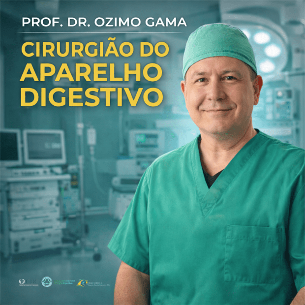
Ozimo Pereira Gama Filho
Adjunct Professor at the Federal University of Maranhão
No conflict of interest
- INTRODUCTION
- HISTORICAL ASPECTS
- ANATOMICAL FUNDAMENTALS
- INDICATIONS
- TYPES & TECHNIQUES
- ADVERSE EVENTS
- CUSTO RATIO x EFFECTIVENESS
- CONCLUSIONS
SUMMARY
In the last decade, the use of gastrostomies has been widely indicated as the preferred form of access to the gastrointestinal tract for feeding in chronic conditions and during recovery from acute conditions such as trauma. Together with this increase in indications, new techniques have been developed that have made gastrostomies simpler and less risky. From the classical technique of Stamm performed by laparotomy, two new alternatives that do not require laparotomy emerged: percutaneous endoscopic gastrostomy (PEG) and fluoroscopy gastrostomy. Its main benefit is to avoid a laparotomy, with less associated postoperative pain and earlier return of gastrointestinal function. Although peg is currently widely accepted as the insertion technique of choice due to its simplicity and efficacy, there are patients who are not candidates for an endoscopic approach. In this article we seek to clarify the indications, technical aspects and perioperative care of patients undergoing gastrostomy.
Keywords: Surgical Procedures; Ostomies; Gastrostomy.
Area of Knowledge: General Surgery
- INTRODUCTION
The main indication for enteral or parenteral feeding in the perioperative period is the provision of nutritional support to supply the metabolism of patients with inadequate oral intake. Enteral feeding is the preferred method in relation to parenteral feeding in patients with gastrointestinal dysfunction in the perioperative period due to the inherent risks associated with parenteral nutritional support, such as: infectious complications of the access routes, higher operational cost, and the inability to parenteral nutrition to provide adequate enteral stimulation and subsequent involvement of the intestinal defense barrier [1,2]. In addition, enteral feeding may decrease the risk of bacterial translocation and corresponding bacteremia [3]. Gastric nutritional support is the most common type used. Access to insert the gastrostomy probe can be achieved using endoscopy, interventional radiologia, or surgical techniques (open or laparoscopic). However, since its description in the 1980s [4], percutaneous endoscopic gastrostomy (PEG) is currently considered the method of choice for medium and long-term enteral support.
1.1 Objective: This article reviews the current knowledge about GOSTROSTOMIA in the medical literature, emphasizing the technical and perioperative aspects.
- HISTORICAL ASPECTS
In 876, Verneoil [5] successfully made the first gastrostomy in humans. Since then, several technical modifications have been suggested, such as witzel’s technique in 1891, in which a subseroso tunnel is made on the probe [6]. Stamm, in 1894 [7], described one of the most performed techniques today and in the history of surgical gastrostomy, which consists in the making of suture in a pouch to invaginate the probe inserted into the stomach [8]. In 1980, percutaneous endoscopic gastrostomy was described by Gauderer et al. [4] , which transformed the technique of making gastrostomy.
- ANATOMICAL FUNDAMENTALS
The stomach is a J-shaped dilated cylindrical organ that rests in the left epigastric and hypochodrial region of the abdomen at the level of the first lumbar vertebra. It is previously limited by the left hemidiaphragm, the left lobe of the liver and a triangular portion of the anterior abdominal wall. Subsequently, the pancreas, left kidney and adrenal delimit the stomach. The spleen is posterolaterally and the transverse colon is inferior. It is fixed at two points of continuity: gastroesophageal, superiorly and the duodenal, retroperitoneally. Its ligament attachments also help you in fixation to adjacent organs: gastrophemic (diaphragm), hepatogastric or minor omentum (liver), gastrosplenic or gastrolienal (spleen), and gastrocholic or omentum major (transverse colon). The anatomical regions of the stomach can be distinguished as this: começa superiorly in the continuity of the abdominal part of the esophagus and dthe gastroesophageal junction, the cardiac part of the stomach. Soon below this portion, lies the bottom of the stomach that expands to the left extending above thegastroesophageal junction, forming an acute angle with the distal esophagus known as cardiac notches. The body s andextends as a distensible reservoir and forms a medial edge called the smallest curvature to the right and a side edge called the largest curvature on the left. The gastric den of the stomach is not anatomically distinguishable, but it is estimated to be a region of the angular isis along the distal minor curvature to a point along a lower line to the distal major curvature. It thus ends bymouthing r into the pyloric canal limited by the pyloric sphincter, a palpable thickened ring of muscle that is continuous with the first part of the duodenum.
- INDICATIONS
Gastrostomy is used in the following situations:
- Gastric decompression: can be obtained by means of temporary gastrostomy, occasionally recommended, as a complement to large abdominal operations for which gastric stems, prolonged “adynamic ileus” and digestive fistulas are foreshadowed.
- Nutritional Support: b.1 Temporary; indicated when access to the digestive tract is temporarily impaired for recovery and maintenance of nutritional status (E.g. CEsophageal EC); b.2 Definitive; as palliative therapy in patients with unresectable malignant neoplasia of the head and neck, as wellas n degenerative neurological diseases that lead to irreversible disorders of deglutition.
However, the decision to perform a gastrostomy, as well as its route (surgical, radiological or endoscopic) should be individualizedaccording to the needs, diagnosis, life expectancy of the patient and the available hospital logistics. The objective is not only to optimize perioperative recovery, to improve survival and nutritional status of the patient, but also to promote quality of life, which is not necessarily correlated with nutritional improvement only [9]. Therefore, the appropriate indication, like any other surgical intervention, must be clearly establishedand informed before it is performed. Some of the absolute contraindications of gastrostomy are summarized in Table 1. In addition to absolute contraindication conditions, other situations such as the presence of non-obstructive oromyctological oresophageal malignancy, hepatomegaly, splenomegaly, peritoneal dialysis, portal hypertension with gastric varicose veins, and a history of partial gastrectomy are also considered relative contraindications.
| ABSOLUTE CONTRA – INDICATIONS |
| Coagulopatia Severa (INR > 5, Plaquetas < 50.000 e TPT > 50s) |
| Hemodynamic Instability |
| Septic Shock |
| Refractory Ascites |
| Peritonitis |
| Dermatological infection in the upper abdomen |
| Carcinomatose Peritoneal |
| Interposition of organs that prevent gastric access |
| History of Total Gastrectomy |
| Stenosis or Pyloric Obstruction |
| Severe gastroparesis, in cases of indication for nutritional support |
| Absence of Informed Consent |
- TYPES & TECHNIQUES
Currently there are three techniques for performing gastrostomy: radiological, through percutaneous gastrostomy by fluoroscopy, percutaneous endoscopic gastrostomy (PEG) and surgical gastrostomy. Due to the didactic characteristics of this material, we will focus on endoscopic and surgical gastrostomy , which becomes the main option in the following situations: 1) when the patient will already undergo a laparotomy due to some abdominal condition ; 2) impossibility of performing gastroscopy to perform gastrostomy endoscopic percutaneous (PEG) ; 3) in case of peg technical failure; 4) unavailability of resources for the preparation of PEG or percutaneous gastrostomy by fluoroscopy.
- ENDOSCOPIC PERCUTANEOUS GASTROSTOMY
The informed consent form must be obtained from patients or their legal representatives. Patients should fast for a minimum of 8 hours and receive prophylactic antibiotics one hour before proceeding and intravenous administration of 1-2 g of cefazolin is recommended. The technique introduced by Gauderer et al [4] is the most used technique to insert the PEG gastrostomy probe. In this method, a guide wire is used, inserted in the distal gastric chamber through a needle puncture n to the anterior abdominal wall. This guide wire is then seized endoscopically with a handle and then removed through the esophagus and mouth. Subsequently, the guide wire is fixed to the end of the gastrostomy probe and then pulled from the mouth to the esophagus, stomach and then out to the abdominal wall, where it will be fixed.
- SURGICAL GASTROSTOMY
Surgical gastrostomy can be performed in two ways: 1) via laparotomy – the predominant form; and 2) laparoscopic approach. The preparations are the same as the endoscopic pathway.
5.2.1. GASTROSTOMIA At STAMM
After adequate asepsis and antisepsis, with the patient under anesthesia and in horizontal dorsal decubitus, the technical steps are as follows: 1. Median laparotomy (supraumbilical median incision); 2. Identification of the gastric body; 3. Stomach hold with Babcock tweezers (to evaluate the approach of the stomach to the peritoneum); 4. Suture in pouch (circular area of 2cm) – atraumatic absorbable thread; 5. Section of the stomach wall (0.5cm) – (incision with scalpel or Electrocautery in the center of the suture, of sufficient size, for the placement of a probe with 20 to 26 French); 6. Placement of the Gastrostomy probe in the extension of 5-6 cm, followed by suture closure in a pouch); 7. Tie the suture threads in a pouch around the probe; 8. Apply a second suture in a pouch 1cm above the first (seromuscular stitches); 9. Externalization of the probe by counter opening on the left flank; 10. Fix the stomach wall to the abdominal wall in 4 cardinal points (external ration with the two Kocher tweezers used for grip of the alba line and against traction by means of the index fingers of the wall of the left hipochondrio to approach the parietal peritoneum of the gastric wall); 11. Fixation of the probe to the skin (point with nonabsorbable wire); 12. Closure of the abdominal wall (synthesis of the wall with approximation of the alba thread by continuous suture with monofilament thread 1-0 or 2-0 and of the skin with separate points of nylon 3-0); 13. Dressing.
5.2.1 GASTROSTOMIA EM WITZEL
The initial technical steps from gastrostomy to Witzel are like those of gastrostomy to Stamm, including fixation of the probe to the stomach by a pouch suture. Then, the probe is placed on the gastric wall and a tunnel of 8-10 cm is made by seromuscular suture (continuous or with separate points of absorbable or nonabsorbable thread) covering it and externalization is performed by counteropening.
- ADVERSE EVENTS
According to the literature, the rate of complications for different procedures varies due to the heterogeneity of the samples evaluated. For surgical gastrostomy, the reported complication rates are between 1% and 35%, while for percutaneous radiological gastrostomy it is 3% to 11%, and for percutaneous endoscopic, 17%–32%, the main related adverse event is surgical site infection [10, 11]. Although considered a basic procedure, gastrostomy is associated with an extensive list of related technical complications, care and use of the probe. Serious problems related to the technique include separation of the stomach from the abdominal wall (leading to peritonitis), separation of wounds, hemorrhage, infection, lesion of the posterior gastric wall or other organs, and placement of the tube in an inappropriate place of gastric position. Separation of the stomach from the abdominal wall usually occurs due to inadvertent and premature displacement of the tube, particularly with balloon-like devices, or a rupture during a catheter change. It requires immediate attention, being treated with laparotomy, although in selected cases laparoscopic correction is possible. Most complications can be avoided with the careful choice of the type of procedure, from the appropriate ostomy device, considering it an important intervention and using meticulous technique with the proper approximation of the stomach to the abdominal wall and outflow of the probe through a counter-incision (in conventional procedures), thus avoiding probes in the midline or awfully close to the costal edge.
- COST VS. EFFECTIVENESS
A recent study [12] compared the cost associated with the different gastrostomy techniques, and the results of the evaluation showed variable the benefits of each of the individual percutaneous procedures, indicating that surgical gastrostomy was the onerous mais of the three modalities due to higher costs, complications, and recovery time, as well as the endoscopic technique presenting the cost effectiveness ratio.
- CONCLUSIONS
Despite the technique employed, the decision to performa gastrostomy is not based only on the patient’s survival expectancy, because the adequate indication provides a better quality of life even when the survival of the patient after the procedure is severely limited. Therefore, understanding of techniques, indications, complication rates is essential to guide the surgical team in the scope of multidisciplinary care, as well as the education of patients and their caregivers is vital to ensure the correct maintenance of the devices, thus ensuring adequate nutritional intake of the patient and minimizes complication rates.
References
1 Alverdy J, Chi HS, Sheldon GF. The effect of parenteral nutrition in gastrointestinal immunity. The importance of de estimulação enteral. Ann Surg, 1985; 202: 681-684 [PMID:3935061]
2 Deitch EA, Ma WJ, Ma L, Berg RD, Specian RD. Protein malnutrition predisposes to inflammation-induced intestinal origin septic states. Ann Surg, 1990; 211: 560-567; discussion 560-567 [PMID: 2111125]
3 Deitch EA, Winterton J, Li M, Berg R. The intestine as a portal of entry to bacteremia. Role of protein malnutrition. Ann Surg 1987; 205: 681-692 [PMID: 3592811]
4 Gauderer MW, Ponsky JL, Izant RJ. Gastrostomia sem laparotomy: percutaneous endoscopic technique. J Pediatrician Surg, 1980; 15: 872-875 [PMID: 6780678]
5 Anselmo CB, Tercioti Júnior V, Lopes LR, Coelho Neto JS, Andreollo NA. Surgical gastrostomy: current indications and complications in patients of a university hospital. Rev Col Bras Cir. [Internet journal] 2013;40(6). Available in URL: http://www.scielo.br/rcbc
6 Witzel O. For gastric fistula technique. Chir Zbl. 1891;18:601-4.
7 Stamm M. Gastrostomy: a new method. Med News. 1894;65:324.
8 JP grant. Comparison of percutaneous endoscopic gastrostomy com gastrostomia strain. Ann Surg. 1988;207(5):598-603
9 Bannerman E, Pendlebury J, Phillips F, Ghosh S. Cross-sectional and longitudinal study of health-related quality of life after percutaneous gastrostomy. Eur J Gastroenterol Hepatol 2000; 12: 1101-1109 [PMID: 11057455]
10 Möller P, Lindberg CG, Zilling T. Gastrostomy by various techniques: evaluation of indications, outcome and complications. Scand J Gastroenterol. 1999;34(10):1050-4.
11 Clarke E, Pitts N, Latchford A, Lewis S. A major prospective audit of morbidity and mortality associated with food gastrostomies in the community. Clin Nutr. 2017 Apr;36(2):485-490. DOI: 10.1016/j.clnu.2016.01.008. EPub 2016 January 21. PMID: 26874913.
12 Wollman B, D’Agostino HB, Walus-Wigle JR, Easter DW, Beale A (1995) Radiological, endoscopic and surgical gastrostomy: an institutional evaluation and meta-analysis of the literature. Radiology 197: 699–704.
A Arte da Anatomia (EBook)
Desde a Antiguidade, os médicos, anatomistas e artistas se dedicaram a descrever e representar o corpo humano, por meio de desenhos, pinturas, esculturas e outras formas de representação artística. Com o passar dos séculos, houve uma evolução significativa na forma como as ilustrações anatômicas eram produzidas, desde as primeiras representações rudimentares até as ilustrações altamente detalhadas e realistas que temos hoje.
Boa Leitura!!!
Michelangelo Buonarroti
O Anatomista Renascentista
“A MEDICINA é a arte de manter a saúde e a vida, e a ARTE é a Medicina da alma.” – Platão
Michelangelo Buonarroti (1475-1564), um dos maiores mestres da arte ocidental, é amplamente conhecido por suas impressionantes esculturas e afrescos. Entretanto, sua contribuição para o estudo da anatomia humana também merece destaque. O genial artista florentino não se limitou apenas a criar obras de arte, mas também se dedicou ao estudo meticuloso do corpo humano, revelando uma compreensão que transcendeu sua época. A interseção entre arte e ciência é um tema fascinante, especialmente quando se considera a figura de Michelangelo Buonarroti, um dos maiores gênios da Renascença. Embora seja amplamente reconhecido por suas magníficas esculturas e afrescos, como o famoso “Davi” e a Capela Sistina, a profundidade de seu conhecimento anatômico é frequentemente subestimada. Este post explora como Michelangelo não apenas revolucionou a arte, mas também contribuiu para o entendimento da anatomia humana, influenciando práticas médicas e cirúrgicas que perduram até hoje.
Introdução
Nascido em uma família modesta em Florença, Michelangelo começou sua trajetória artística muito jovem. A fama chegou cedo com obras como a “Pietà” e o “David”, e seus afrescos na Capela Sistina continuam a ser um marco na arte ocidental. No entanto, menos conhecido é seu trabalho em anatomia, um campo que ele explorou de forma intensiva para aprimorar sua técnica artística e contribuir para o conhecimento médico da época. Nascido em 1475, Michelangelo começou sua formação artística sob a orientação de Domenico Ghirlandaio e Bertoldo di Giovanni. Desde jovem, ele se interessou pela anatomia, realizando dissecações de cadáveres no hospital do Mosteiro de Santo Spirito, em Florença. Essa prática, embora controversa na época, permitiu que ele adquirisse um conhecimento profundo da estrutura e função do corpo humano, que se refletiu em suas obras
Estudos Anatômicos
No início dos anos 1500, quando Michelangelo tinha cerca de 25 anos, ele iniciou seus estudos anatômicos. Em uma época em que a dissecação de cadáveres humanos era proibida pela Igreja Católica, Michelangelo obteve corpos de condenados à morte através do Hospital de Santa Maria Nuova, em Florença. A dissecação era permitida para fins de ensino médico em Florença, o que proporcionou a Michelangelo a oportunidade de estudar o corpo humano com detalhes.
A arte renascentista era marcada por um desejo de representar o corpo humano de maneira realista. Michelangelo, assim como seus contemporâneos, estudou a anatomia com rigor. O artista acreditava que “os membros da arquitetura dependem dos membros do homem”, enfatizando a importância do conhecimento anatômico para a criação artística. Ele fez moldes de músculos em diversas posturas, aplicando esse conhecimento em suas representações de figuras nuas, como os “ignudi” na Capela Sistina.
Michelangelo realizou suas dissecações em segredo, utilizando uma sala alugada próxima ao hospital e contando apenas com um ajudante de confiança. Ele estudou o corpo humano por aproximadamente 18 meses, realizando pelo menos duas dissecações completas: uma de um homem e outra de uma mulher. Durante esse período, fez centenas de desenhos e anotações detalhadas dos órgãos, ossos e músculos, proporcionando uma visão profunda e detalhada da anatomia humana.
Trabalhos Anatômicos
O conhecimento anatômico de Michelangelo não se limitou à arte; suas observações e dissecações influenciaram o entendimento da anatomia humana na medicina. Ele estudou não apenas a forma, mas também os movimentos e posturas do corpo, o que é essencial para cirurgiões e profissionais de saúde. As descrições detalhadas de músculos e ossos em suas obras oferecem insights valiosos que podem ser aplicados no campo cirúrgico, especialmente em procedimentos que envolvem a manipulação de estruturas musculoesqueléticas. Apesar de ter deixado poucos trabalhos anatômicos concluídos, os desenhos de Michelangelo são valiosos para a história da anatomia. Entre as principais obras anatômicas que ele produziu estão:
- Estudo para a Leda e o Cisne – Um desenho a carvão que representa uma figura feminina em uma pose que permite uma visualização detalhada da musculatura das costas, braços e pernas. Este estudo evidencia a habilidade de Michelangelo em capturar a complexidade dos músculos humanos.
- Estudo para o Braço Direito da Leda e o Cisne – Outro desenho a carvão que foca na musculatura do braço direito da figura feminina, mostrando a precisão com que Michelangelo abordava a anatomia dos membros.
- Desenho da Cabeça de Lutador – Um esboço que detalha a anatomia da cabeça e do pescoço de um lutador, revelando a complexidade dos músculos e tendões.
- Anatomia dos Músculos da Perna – Um desenho que exibe a musculatura da perna em diferentes ângulos, com atenção especial aos músculos da panturrilha, demonstrando a habilidade de Michelangelo em representar a anatomia com precisão.
- Anatomia da Cabeça – Uma série de desenhos que abordam diferentes aspectos da anatomia da cabeça, incluindo a musculatura facial e o crânio.
- Anatomia do Braço – Outra série de desenhos que mostram a musculatura do braço em variados ângulos, com destaque para os músculos do antebraço.
A Anatomia na Arte de Michelangelo
Michelangelo utilizou seu conhecimento anatômico para enriquecer suas obras artísticas, proporcionando uma representação extremamente realista do corpo humano. Sua habilidade em retratar músculos, veias e ossos com grande precisão não só embelezou suas esculturas, mas também demonstrou um profundo entendimento da anatomia. Michelangelo frequentemente incorporava elementos anatômicos em suas pinturas e esculturas de maneira que muitas vezes passavam despercebidos. Por exemplo, a forma do manto de Deus na Capela Sistina tem sido interpretada como uma representação do rim humano, enquanto a figura de São Bartolomeu no “Juízo Final” exibe uma pele esfolada que remete a dissecações. Essas representações não apenas demonstram seu domínio técnico, mas também refletem uma compreensão profunda da anatomia que era incomum para a época.
O Legado de Michelangelo
A influência de Michelangelo na medicina e na arte é inegável. Ele não apenas elevou a escultura e a pintura a novos patamares, mas também ajudou a estabelecer uma base para o estudo da anatomia que continuaria a evoluir nos séculos seguintes. Sua abordagem holística, que unia arte e ciência, continua a inspirar artistas e médicos contemporâneos.
Músculos e Veias
Michelangelo foi um mestre em retratar a musculatura e as veias com uma precisão que parecia quase sobre-humana. Em suas esculturas, como o “David”, ele conseguiu capturar a aparência dos músculos e veias de forma tão realista que quase parece que eles estão prestes a saltar da pedra. Seu uso detalhado da musculatura para transmitir força e vitalidade é um exemplo notável de como a arte pode ser usada para explorar a complexidade da forma humana.
Ossos e Órgãos Internos
A compreensão de Michelangelo da estrutura óssea e dos órgãos internos é igualmente impressionante. Em suas obras, ele retratou os ossos com uma precisão detalhada, especialmente notável na escultura “David”. Seus desenhos anatômicos mostram um entendimento profundo dos órgãos internos, como o coração e os pulmões, que eram detalhadamente representados e contribuiram para o conhecimento médico da época.
Detalhes Ocultos
Além das representações evidentes, Michelangelo também incluiu detalhes anatômicos mais sutis e ocultos em suas obras. Um exemplo é a escultura “Moisés”, onde ele esculpiu uma protuberância sob a barba do personagem que alguns acreditam ser uma nodulação, visível apenas com modernas técnicas de análise de imagem. Essas observações demonstram a atenção meticulosa de Michelangelo aos detalhes anatômicos, indo além do que era visível a olho nu.
Legado
A dedicação de Michelangelo ao estudo da anatomia não só aprimorou suas habilidades artísticas, mas também fez contribuições significativas ao conhecimento médico. Seus desenhos anatômicos e sua abordagem detalhada da estrutura humana abriram novos caminhos para a compreensão da forma humana e tiveram um impacto duradouro na arte e na Medicina. Sua abordagem interdisciplinar mostra como a arte e a ciência podem se entrelaçar para expandir nosso entendimento do corpo humano e melhorar a prática médica. Michelangelo nos deixou um legado que continua a influenciar tanto artistas quanto médicos, demonstrando que a busca pelo conhecimento é uma jornada que transcende disciplinas.
“A anatomia é a ciência que nos ensina a conhecer a natureza do homem, e é indispensável para quem quer entender a arte de curar.” – Hippocrates, médico grego.
Conclusão
Michelangelo Buonarroti não foi apenas um artista extraordinário, mas também um anatomista renascentista cuja compreensão da estrutura humana foi avançada para sua época. Seus estudos anatômicos não apenas enriqueceram suas obras de arte, mas também contribuíram para o desenvolvimento do conhecimento médico. O impacto de seu trabalho é um exemplo brilhante de como a paixão pela arte e pela ciência pode gerar descobertas significativas e inspiradoras.
Gostou? Nos deixe um comentário ✍️, compartilhe em suas redes sociais e/ou mande sua dúvida pelo 💬 Chat On-line em nossa DM do Instagram.
Hashtags
#Michelangelo #AnatomiaRenascentista #HistóriaDaArte #EstudosAnatômicos #AvançosNaMedicina